HARIBOSS logo

HARIBOSS

Harnessing RIBOnucleic acid - Small molecules Structures

RNA-SM complexes

× Close

Combine filters to refine your query. Each filter opens a dialog showing the values available in the data.


270 RNA-SM complexes found
Structure of complex 4x4v

4x4v

Crystal structure of the A.fulgidus CCA-adding enzyme in complex with a human MenBeta minihelix ending in CCACC and AMPcPP

SM: 5GP 5GP 5GP APC APC

Structure of complex 4ybb

4ybb

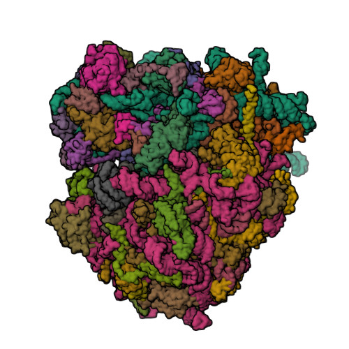
High-resolution structure of the Escherichia coli ribosome

SM: 1PE 1PE PG4 PG4 PG4 PG4 PG4

Structure of complex 5dhc

5dhc

Cooperativity and Downstream Binding in RNA Replication

SM: 5GP 5GP 5GP 5GP 5GP 5GP 5GP 5GP

Structure of complex 5hbx

5hbx

RNA primer-template complex with 2-methylimidazole-activated monomer analogue-2 binding sites

SM: PZG PZG PZG PZG PZG PZG PZG PZG

Structure of complex 5hby

5hby

RNA primer-template complex with 2-methylimidazole-activated monomer analogue-3 binding sites

SM: PZG PZG

Structure of complex 5hkv

5hkv

The crystal structure of the large ribosomal subunit of Staphylococcus aureus in complex with lincomycin

SM: 3QB

Structure of complex 5it8

5it8

High-resolution structure of the Escherichia coli ribosome

SM: 1PE 1PE PG4 PG4 PG4 PG4 PG4

Structure of complex 5j5b

5j5b

Structure of the WT E coli ribosome bound to tetracycline

SM: 1PE 1PE PG4 PG4 PG4 PG4 PG4 TAC TAC

Structure of complex 5j7l

5j7l

Structure of the 70S E coli ribosome with the U1052G mutation in the 16S rRNA bound to tetracycline

SM: 1PE 1PE PG4 PG4 PG4 PG4 PG4 TAC TAC TAC TAC

Structure of complex 5j88

5j88

Structure of the E coli 70S ribosome with the U1060A mutation in 16S rRNA

SM: 1PE 1PE PG4 PG4 PG4 PG4 PG4

Structure of complex 5j8a

5j8a

Structure of the E coli 70S ribosome with the U1052G mutation in 16S rRNA bound to tigecycline

SM: 1PE 1PE PG4 PG4 PG4 PG4 PG4 T1C T1C

Structure of complex 5j91

5j91

Structure of the Wild-type 70S E coli ribosome bound to Tigecycline

SM: 1PE 1PE PG4 PG4 PG4 PG4 PG4 T1C T1C

Structure of complex 5jc9

5jc9

Structure of the Escherichia coli ribosome with the U1052G mutation in the 16S rRNA

SM: 1PE 1PE PG4 PG4 PG4 PG4 PG4

Structure of complex 5ndg

5ndg

Crystal structure of geneticin (G418) bound to the yeast 80S ribosome

SM: GET GET GET GET GET GET GET GET GET GET GET GET GET GET GET GET GET GET GET GET GET

Structure of complex 5ndv

5ndv

Structure of complex 5obm

5obm

Crystal structure of Gentamicin bound to the yeast 80S ribosome

SM: LLL LLL LLL LLL LLL LLL LLL LLL LLL LLL LLL LLL LLL LLL LLL LLL LLL LLL LLL LLL LLL LLL LLL LLL LLL LLL LLL LLL LLL LLL LLL LLL LLL LLL LLL LLL LLL LLL LLL LLL LLL LLL LLL LLL LLL LLL LLL LLL LLL LLL LLL LLL LLL LLL LLL LLL LLL LLL LLL LLL LLL LLL LLL LLL LLL LLL LLL LLL LLL

Structure of complex 5tgm

5tgm

Crystal structure of the S.cerevisiae 80S ribosome in complex with the A-site bound aminoacyl-tRNA analog ACCA-Pro

SM: PHE PHE SPS SPS

Structure of complex 5v0j

5v0j

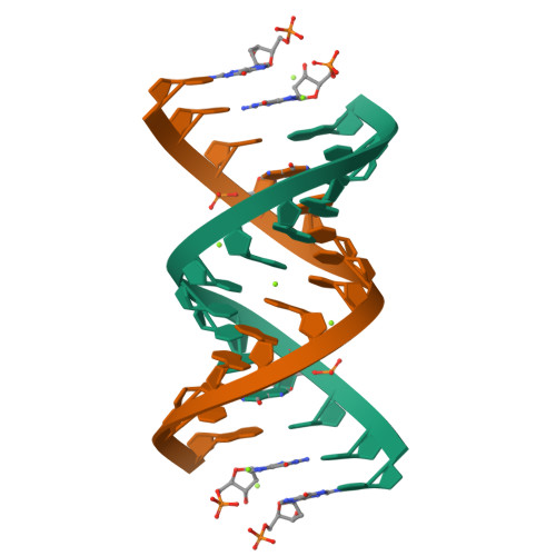
RNA duplex with 2-MeImpG analogue bound-2 binding sites

SM: 8OS 8OS 8OS 8OS

Structure of complex 5v0k

5v0k

RNA duplex with 2-MeImpG analogue bound-3 binding sites

SM: 8OS 8OS

Structure of complex 5vj9

5vj9

Guanidine-II riboswitch P2 hairpin dimer from Pseudomonas aeruginosa

SM: SPM SPM SPM SPM SPM

Structure of complex 5vjb

5vjb

Guanidine-II riboswitch P2 hairpin dimer with 5-bromoU substitution from Pseudomonas aeruginosa

SM: SPM SPM SPM SPM

Structure of complex 6az3

6az3

Cryo-EM structure of of the large subunit of Leishmania ribosome bound to paromomycin

SM: PAR PAR PAR PAR PAR PAR PAR PAR PAR

Structure of complex 6c8d

6c8d

RNA-dGMP complex with Mg ion

SM: DGP DGP DGP DGP

Structure of complex 6c8i

6c8i

RNA-activated 2-AIpG monomer complex, 5 min soaking

SM: EQ4 EQ4 EQ4 EQ4

Structure of complex 6c8j

6c8j

RNA-activated 2-AIpG monomer complex, 15 min soaking

SM: EQ4 EQ4 EQ4 EQ4

Structure of complex 6cab

6cab

RNA-dGMP complex with Sr ion

SM: 5GP 5GP 5GP 5GP

Structure of complex 6e1t

6e1t

Crystal structure of a class I PreQ1 riboswitch complexed with a synthetic compound 1: 2-[(dibenzo[b,d]furan-2-yl)oxy]ethan-1-amine

SM: HLV MES

Structure of complex 6e8s

6e8s

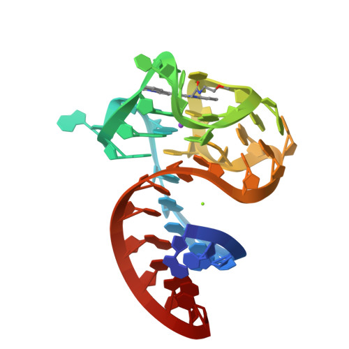
Structure of the iMango-III aptamer bound to TO1-Biotin

SM: EKJ EKJ SPM

Structure of complex 6i1l

6i1l

Crystal structure of FnCas12a in complex with a crRNA guide and ssDNA target

SM: CIT CIT CIT CIT CIT CIT

Structure of complex 6izp

6izp

Solution structure of the complex of naphthyridine carbamate dimer and an RNA with UGGAA-UGGAA pentad

SM: B2R B2R

Structure of complex 6jjh

6jjh

Crystal structure of a two-quartet RNA parallel G-quadruplex complexed with the porphyrin TMPyP4

SM: POH POH

Structure of complex 6p1u

6p1u

Post-catalytic nicked complex of human DNA Polymerase Mu with 1-nt gapped substrate containing template 8OG and newly incorporated CMP

SM: CTP

Structure of complex 6qiq

6qiq

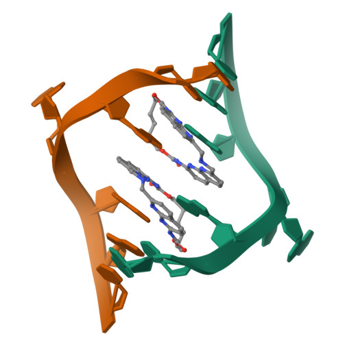
Crystal structure of seleno-derivative CAG repeats with synthetic CMBL3a compound

SM: J48 J48

Structure of complex 6qir

6qir

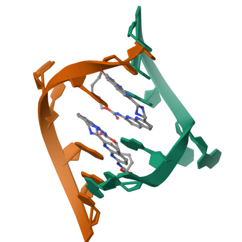
Crystal structure of CAG repeats with synthetic CMBL3a compound (model I)

SM: J48 J48 J48 J48

Structure of complex 6qit

6qit

Crystal structure of CAG repeats with synthetic CMBL3b compound

SM: J48 J48 J48 J48

Structure of complex 6tnu

6tnu

Yeast 80S ribosome in complex with eIF5A and decoding A-site and P-site tRNAs.

SM: 3HE

Structure of complex 6uu3

6uu3

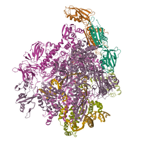
E. coli sigma-S transcription initiation complex with a 4-nt RNA and a CTP ("Old" crystal soaked with GTP, ATP, CTP, and ddTTP for 30 minutes)

SM: D4M

Structure of complex 6uu9

6uu9

E. coli mutant sigma-S transcription initiation complex with an 8-nt RNA ("Fresh" mutant crystal soaked with GTP, UTP, CTP, and ddATP for 30 minutes)

SM: 2DA

Structure of complex 6v9b

6v9b

Co-crystal structure of the fluorogenic Mango-IV homodimer bound to TO1-Biotin

SM: QSA QSA QSA

Structure of complex 6v9d

6v9d

Co-crystal structure of the fluorogenic Mango-IV homodimer bound to TO1-Biotin

SM: QW4 QW4 QW4

Structure of complex 6yl5

6yl5

Crystal structure of the SAM-SAH riboswitch with SAH

SM: SAH SAH SAH SAH SAH SAH SAH SAH SAH

Structure of complex 6ylb

6ylb

Crystal structure of the SAM-SAH riboswitch with SAM

SM: SAM SAM SAM SAM SAM SAM SAM SAM SAM

Structure of complex 6ymi

6ymi

Crystal structure of the SAM-SAH riboswitch with AMP.

SM: AMP AMP AMP AMP AMP AMP AMP AMP AMP CBV CBV

Structure of complex 6ymj

6ymj

Crystal structure of the SAM-SAH riboswitch with adenosine.

SM: ADN ADN ADN ADN ADN ADN ADN ADN ADN

Structure of complex 6ymk

6ymk

Crystal structure of the SAM-SAH riboswitch with AMP

SM: MTA MTA MTA MTA MTA MTA MTA MTA MTA

Structure of complex 6yml

6yml

Crystal structure of the SAM-SAH riboswitch with decarboxylated SAH

SM: AMP DSH DSH

Structure of complex 6ymm

6ymm

Crystal structure of the SAM-SAH riboswitch with SAM from space group P312

SM: SAM SAM SAM

Structure of complex 6ysi

6ysi

Acinetobacter baumannii ribosome-tigecycline complex - 50S subunit

SM: T1C T1C T1C

Structure of complex 6zsa

6zsa

Human mitochondrial ribosome bound to mRNA, A-site tRNA and P-site tRNA

SM: DOL

Structure of complex 7l0z

7l0z

Spinach variant bound to DFHBI-1T

SM: 2ZY SPM SPM SPM

Structure of complex 7l6r

7l6r

Crystal Structure of SARS-CoV-2 Nsp16/10 Heterodimer in Complex with (m7GpppA2m)pUpUpApApA (Cap-1), S-Adenosyl-L-homocysteine (SAH) and Manganese (Mn).

SM: SAH

Structure of complex 7l6t

7l6t

Crystal Structure of SARS-CoV-2 Nsp16/10 Heterodimer in Complex with (m7GpppA2m)pUpUpApApA (Cap-1), S-Adenosyl-L-homocysteine (SAH) and two Magnesium (Mg) ions.

SM: SAH

Structure of complex 7m4v

7m4v

A. baumannii Ribosome-Eravacycline complex: 50S

SM: YQM YQM

Structure of complex 7m4w

7m4w

A. baumannii Ribosome-Eravacycline complex: Empty 70S

SM: YQM YQM YQM

Structure of complex 7m4x

7m4x

A. baumannii Ribosome-Eravacycline complex: P-site tRNA 70S

SM: YQM YQM YQM

Structure of complex 7m4y

7m4y

A. baumannii Ribosome-Eravacycline complex: E-site tRNA 70S

SM: YQM YQM YQM

Structure of complex 7m4z

7m4z

A. baumannii Ribosome-Eravacycline complex: hpf-bound 70S

SM: YQM YQM

Structure of complex 7m8a

7m8a

Human DNA Pol eta S113A with rA-ended primer and dATP: in crystallo reaction for 40 s

SM: DTP

Structure of complex 7m8b

7m8b

Human DNA Pol eta S113A with rA-ended primer and dATP: in crystallo reaction for 140 s

SM: DTP

Structure of complex 7m8c

7m8c

Human DNA Pol eta S113A with rA-ended primer and dATP: in crystallo reaction for 230 s

SM: DTP