Harnessing RIBOnucleic acid - Small molecules Structures
Combine filters to refine your query. Each filter opens a dialog showing the values available in the data.
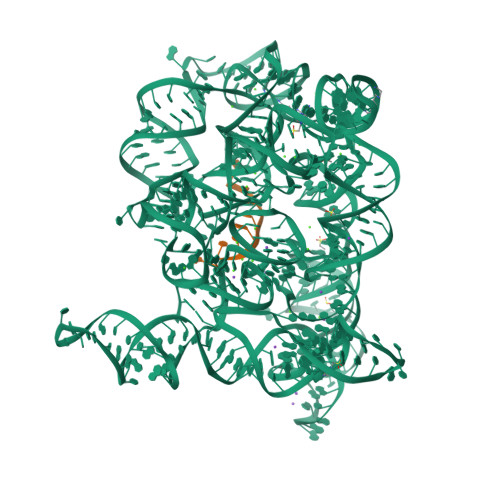
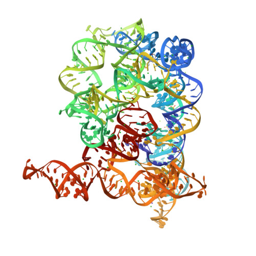
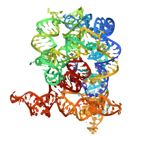
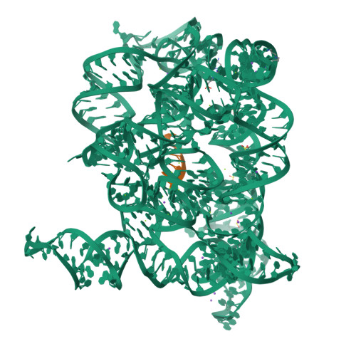
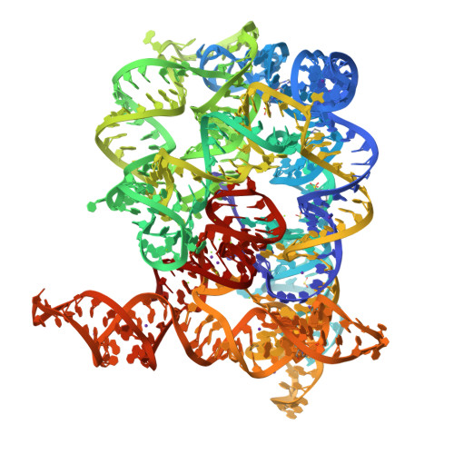
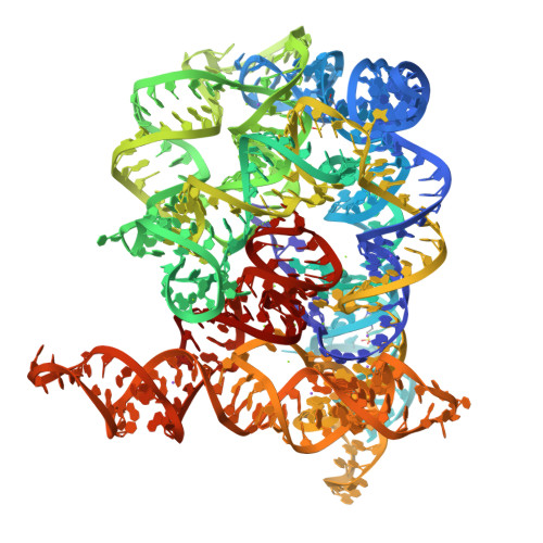
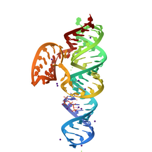
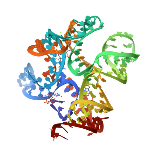
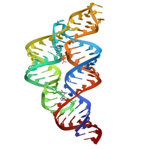
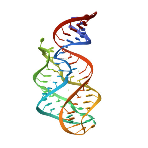
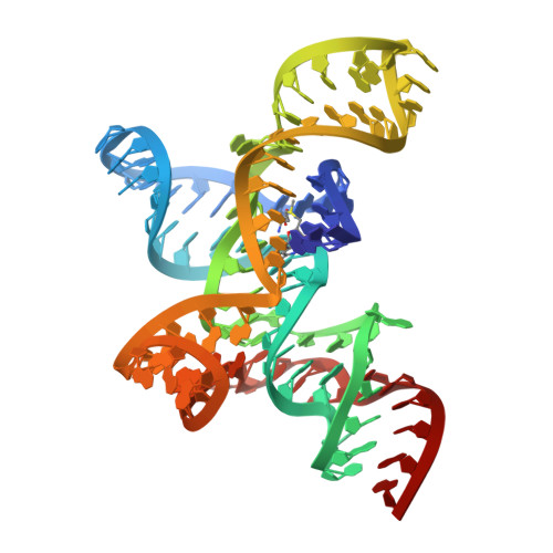
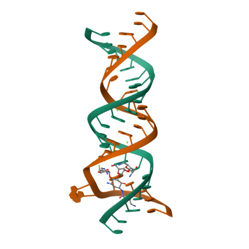
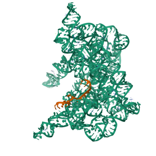
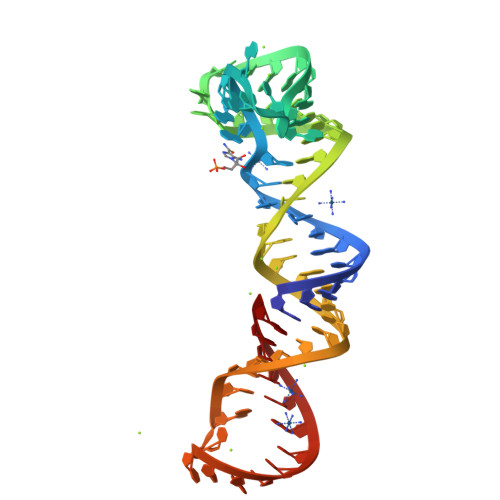
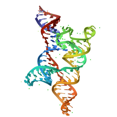
Structure of Oceanobacillus iheyensis group II intron in the presence of K+, Ca2+ and an oligonucleotide fragment substrate (low energy dataset)
SM: EPE EPE EPE EPE EPE SPM SPM
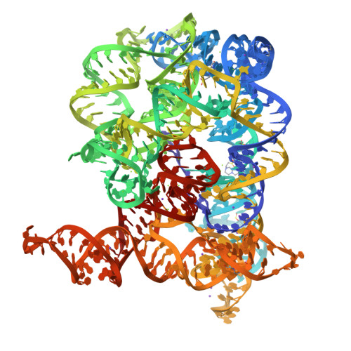
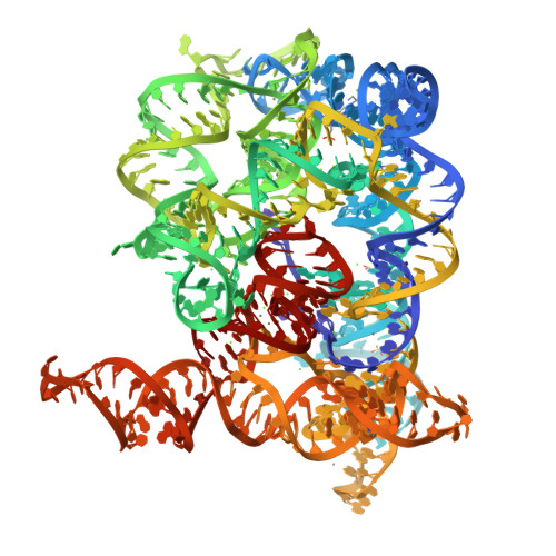
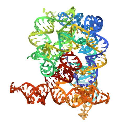
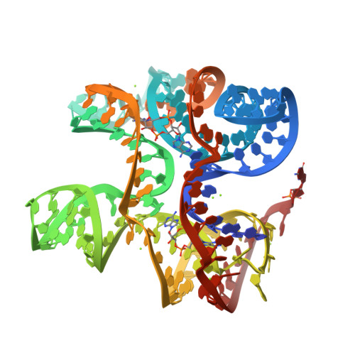
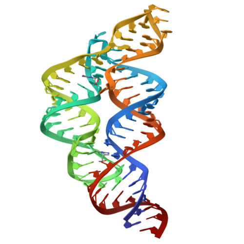
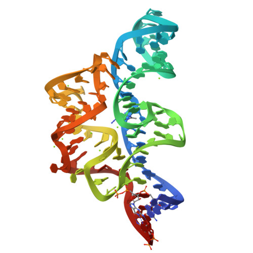
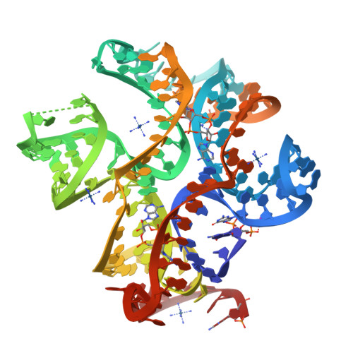
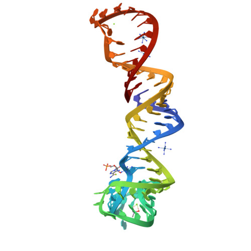
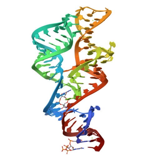
Structure of Oceanobacillus iheyensis group II intron in a ligand-free state in the presence of K+ and Ba2+
SM: EPE EPE EPE EPE
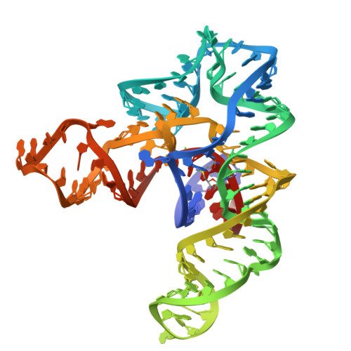
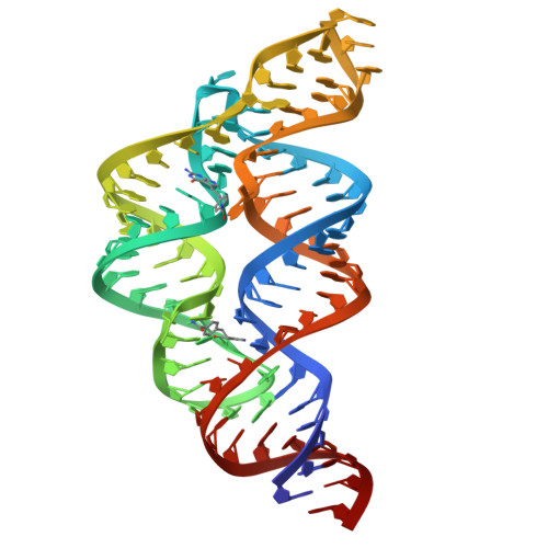
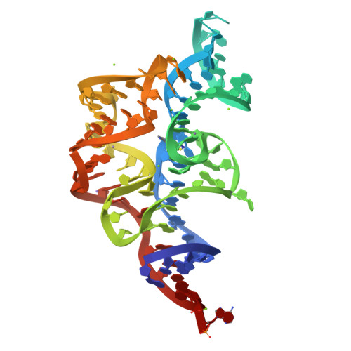
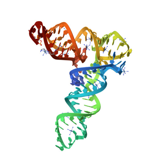
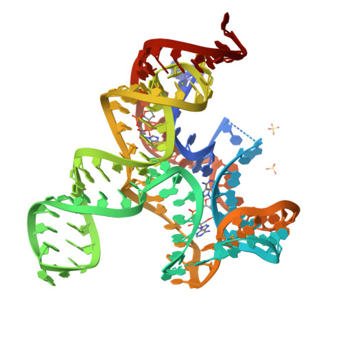
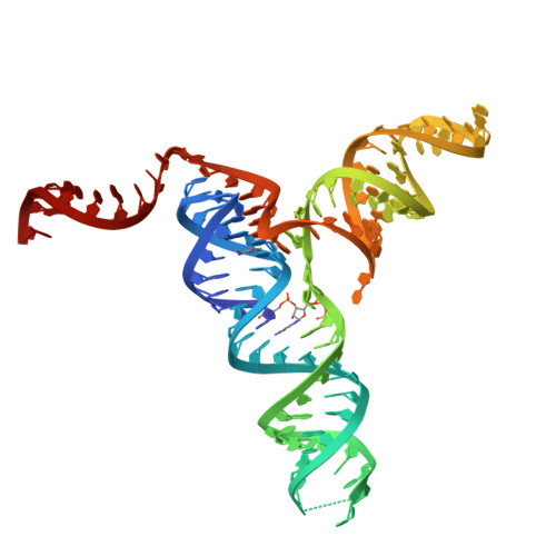
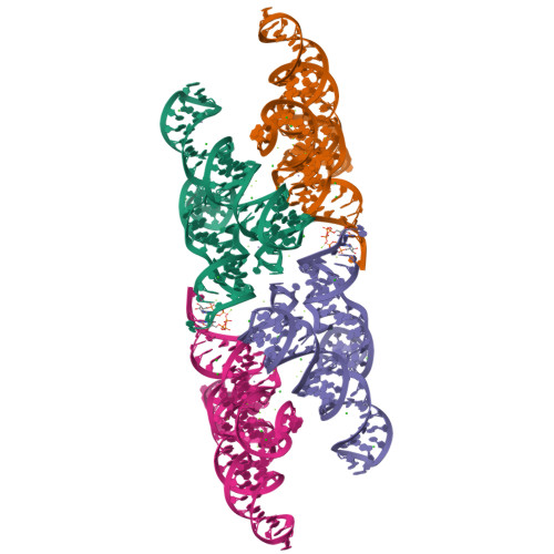
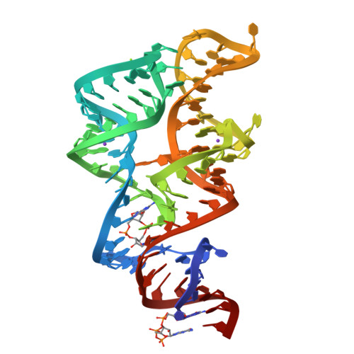
SAM-I riboswitch bearing the H. marismortui K-t-7
SM: SAM
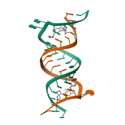
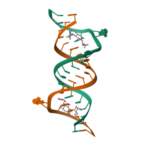
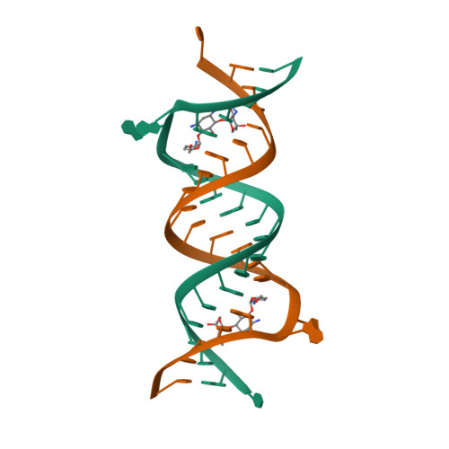
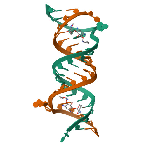
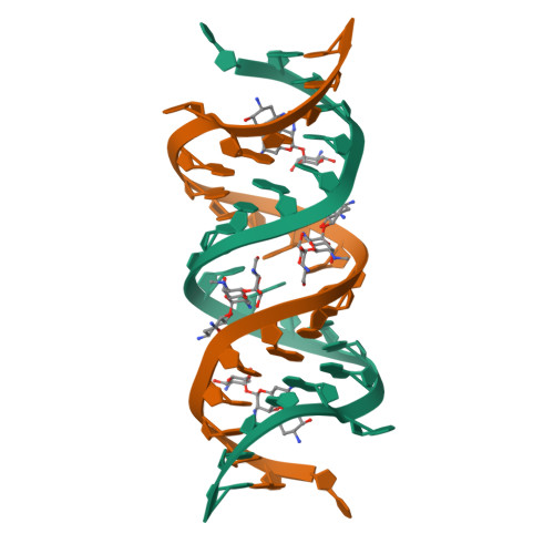
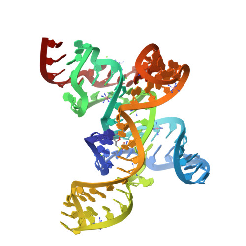
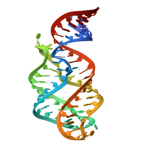
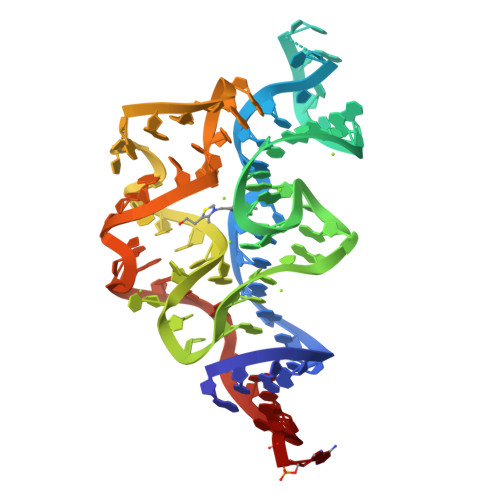
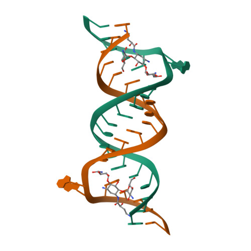
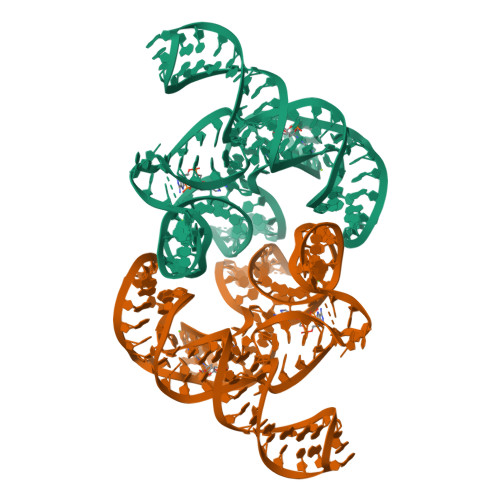
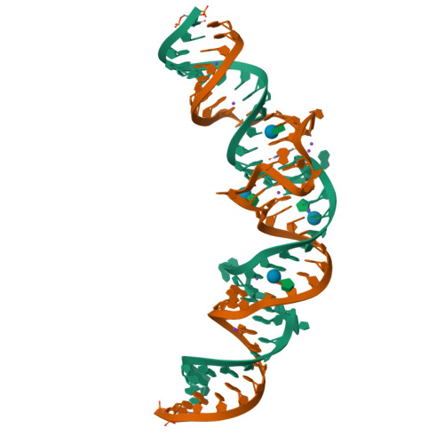
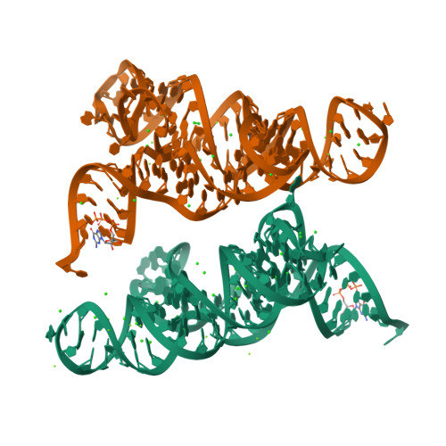
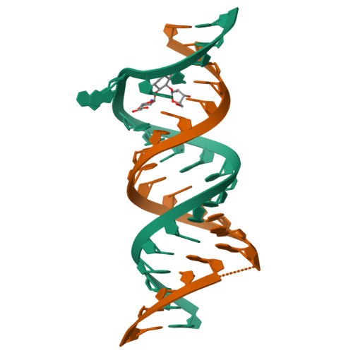
Crystal structure of the bacterial ribosomal decoding site in complex with sisomicin (C2 form)
SM: SIS SIS
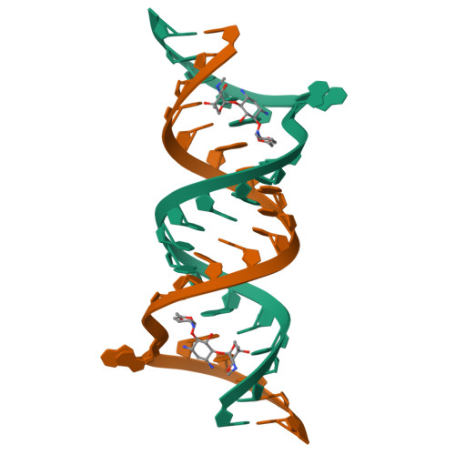
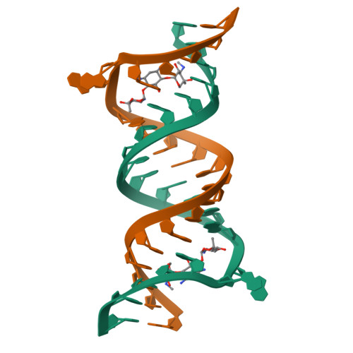
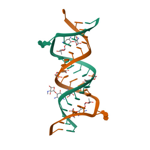
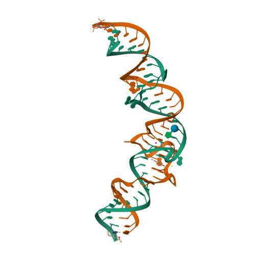
Crystal structure of the bacterial ribosomal decoding site in complex with sisomicin (P21212 form)
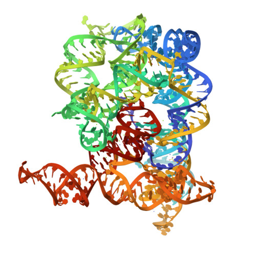
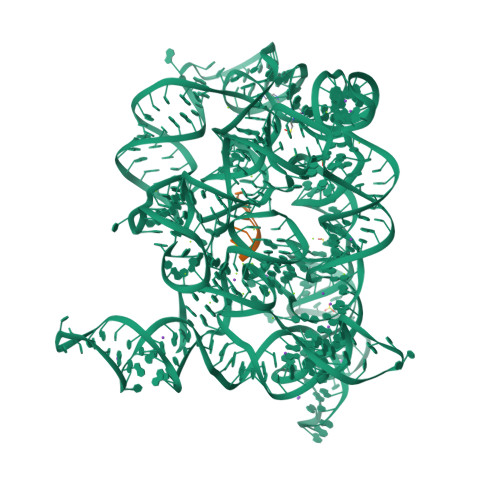
Structure of Oceanobacillus iheyensis group II intron in the presence of K+, Ca2+ and 5'-exon
Structure of Oceanobacillus iheyensis group II intron in the presence of K+, Mg2+ and 5'-exon
SM: EPE EPE EPE SPM SPM
Structure of Oceanobacillus iheyensis group II intron in the presence of K+, Ca2+ and a non-hydrolyzed oligonucleotide substrate
SM: EPE EPE EPE EPE EPE SPM
Structure of Oceanobacillus iheyensis group II intron in a ligand-free state in the presence of K+ and Mg2+
Structure of Oceanobacillus iheyensis group II intron in a ligand-free state in the presence of NH4+ and Mg2+
SM: EPE EPE SPM
Structure of Oceanobacillus iheyensis group II intron in a ligand-free state in the presence of Rb+ and Mg2+
SM: EPE EPE EPE EPE SPM
Structure of Oceanobacillus iheyensis group II intron in a ligand-free state in the presence of Tl+ and Mg2+
SM: EPE EPE EPE SPM
Structure of Oceanobacillus iheyensis group II intron in a ligand-free state in the presence of Cs+ and Mg2+
Crystal structure of the bacterial ribosomal decoding site in complex with 6'-hydroxysisomicin
SM: 6HS 6HS
Structure of Oceanobacillus iheyensis group II intron in the presence of Li+, Mg2+ and 5'-exon
Structure of Oceanobacillus iheyensis group II intron in the presence of K+, Mg2+ and a hydrolyzed oligonucleotide fragment
Structure of Oceanobacillus iheyensis group II intron in a ligand-free state in the presence of Na+ and Mg2+
SM: EPE EPE EPE
Structure of Oceanobacillus iheyensis group II intron C377G mutant in a ligand-free state in the presence of K+ and Mg2+
Crystal structure of the protozoal cytoplasmic ribosomal decoding site in complex with 6'-hydroxysisomicin (P21212 form)
Crystal structure of the protozoal cytoplasmic ribosomal decoding site in complex with 6'-hydroxysisomicin (P212121 form)
Structure of a class II preQ1 riboswitch reveals ligand recognition by a new fold
SM: PRF
Crystal structure of apramycin bound to the leishmanial rRNA A-site
SM: AM2 AM2 AM2 AM2
Crystal structure of geneticin bound to the leishmanial rRNA A-site
SM: GET GET
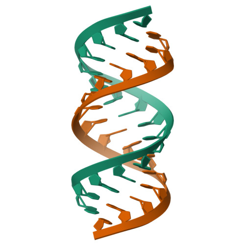
Crystal Structure Analysis of the 16mer GCAGACUUAAGUCUGC
SM: SPM
Thermoanaerobacter pseudethanolicus c-di-AMP riboswitch
SM: 2BA 2BA
Thermovirga lienii c-di-AMP riboswitch
Bacillus subtilis yitJ S box/SAM-I riboswitch
Structure of the SAM-I/IV riboswitch (env87(deltaU92, deltaG93))
Structure of the THF riboswitch
SM: FFO FFO
Structure of the THF riboswitch bound to tetrahydrobiopterin
SM: H4B H4B
Structure of the THF riboswitch bound to pemetrexed
SM: LYA LYA
X-ray crystal structure of the M6" riboswitch aptamer bound to pyrimido[4,5-d]pyrimidine-2,4-diamine (PPDA)
SM: 29G
X-ray crystal structure of the M6C" riboswitch aptamer bound to 2-aminopyrimido[4,5-d]pyrimidin-4(3H)-one (PPAO)
SM: 29H
Crystal structure of the E. coli thiM riboswitch in complex with 5-(azidomethyl)-2-methylpyrimidin-4-amine
SM: 2QB 2QB
Crystal structure of the E. coli thiM riboswitch in complex with (4-(1,2,3-thiadiazol-4-yl)phenyl)methanamine
SM: 2QC
Crystal structure of the E. coli thiM riboswitch in complex with thiamine
SM: VIB
Structure of the SAM-I/IV riboswitch (env87(deltaU92))
Crystal structures of the bacterial ribosomal decoding site complexed with amikacin
SM: AKN AKN
Crystal structure of the bacterial A1408C-mutant ribosomal decoding site in complex with geneticin
SM: GET GET GET
Crystal structure of the tRNA-like structure from Turnip Yellow Mosaic Virus (TYMV), a tRNA mimicking RNA
Crystal structure of the bacterial ribosomal decoding site in complex with 4'-deoxy-4'-fluoro neomycin analog
SM: NMZ
c-di-AMP riboswitch from Thermoanaerobacter pseudethanolicus, iridium hexamine soak
ydao riboswitch binding to c-di-AMP
structure of ydao riboswitch binding with c-di-dAMP
Crystal structure of a eukaryotic group II intron lariat
SM: SPM SPM
Crystal Structure of a PreQ1 Riboswitch
Crystal structure of the Spinach RNA aptamer in complex with DFHBI, barium ions
SM: 38E
Crystal structure of the Spinach RNA aptamer in complex with DFHBI, magnesium ions
Crystal structure of the ZMP riboswitch at 2.50 angstrom
SM: AMZ
Crystal structure of the ZMP riboswitch at 1.80 angstrom
Lactococcus lactis yybP-ykoY Mn riboswitch bound to Mn2+
SM: GTP GTP
Lactococcus lactis yybP-ykoY Mn riboswitch A41U binding site mutant in presence of Mn2+
An Escherichia coli yybP-ykoY Mn riboswitch in the Mn2+-free state
3',3'-cGAMP riboswitch bound with 3',3'-cGAMP
SM: 4BW 4BW
3',3'-cGAMP riboswitch bound with c-di-GMP
SM: C2E C2E
Paromomycin bound to a leishmanial ribosomal A-site
SM: PAR PAR
The structure of A pfI Riboswitch Bound to ZMP
SM: AMZ AMZ
Crystal structure of the Corn RNA aptamer in complex with DFHO, site-specific 5-iodo-U
SM: 747
Crystal structure of the Corn RNA aptamer in complex with DFHO, iridium hexammine soak
Fusobacterium ulcerans ZTP riboswitch bound to ZMP
Current selection range: to
Filter entries " Experimental resolution " between the two values. Global DB range: 0.6 – 11.2
Filter entries " Molecular weight (compound) " between the two values. Global DB range: 150 – 1000
Filter entries " Deposition date " between the two values. Global DB range: 1986 – 2027
Filter entries " Molecular weight (complex) " between the two values. Global DB range: 0 – 13090
Filter entries " Number of atoms " between the two values. Global DB range: 5 – 70
Filter entries " Number of interacting chains " between the two values. Global DB range: 1 – 4
Filter entries " Pocket size " between the two values. Global DB range: 15.9 – 394.0
Filter entries " SiteScore " between the two values. Global DB range: 0.3 – 1.3
Filter entries " Dscore " between the two values. Global DB range: -0.1 – 1.4
Filter entries " Exposure " between the two values. Global DB range: 0.1 – 1.0
Filter entries " Enclosure " between the two values. Global DB range: 0.3 – 1.0
Filter entries " Hydrophobicity " between the two values. Global DB range: 0.0 – 3.1
Filter entries " Hydrophilicity " between the two values. Global DB range: 0.2 – 2.8
Filter entries " Donor/Acceptor " between the two values. Global DB range: 0.0 – 5.0
Filter entries " Volume " between the two values. Global DB range: 21.2 – 1528.8