Harnessing RIBOnucleic acid - Small molecules Structures
Combine filters to refine your query. Each filter opens a dialog showing the values available in the data.
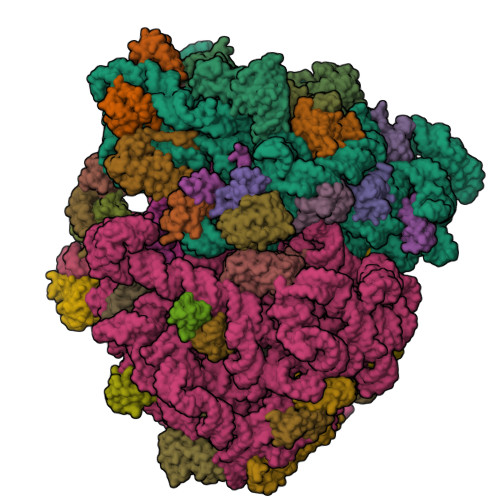
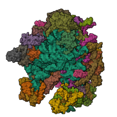
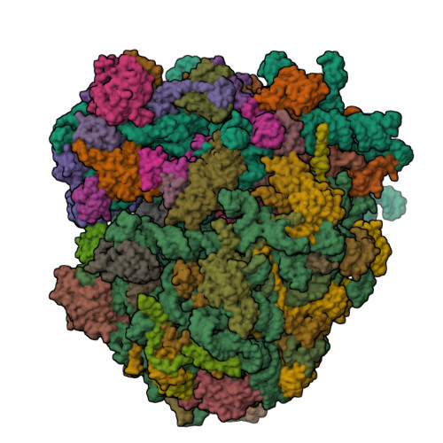
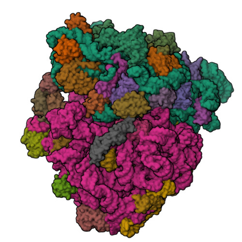
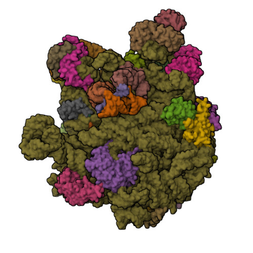
Structure of the Escherichia coli 70S ribosome in complex with EF-Tu and Ile-tRNAIle(LAU) bound to the near-cognate AUG codon (Structure II)
SM: PAR
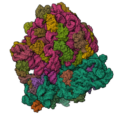
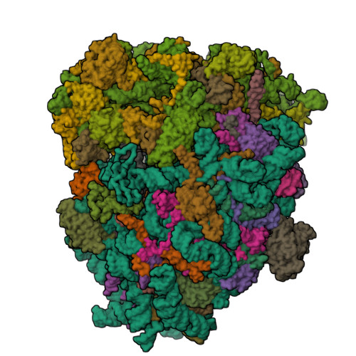
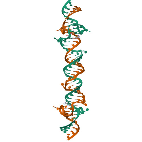
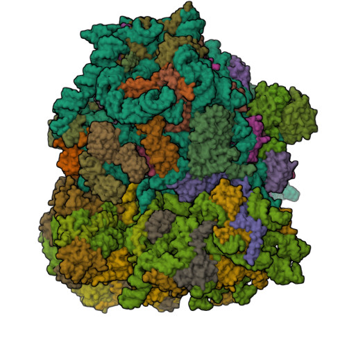
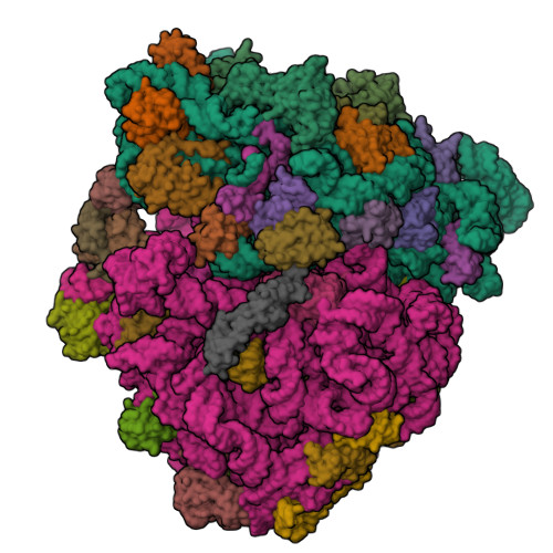
Structure of the Escherichia coli 70S ribosome in complex with A-site tRNAIle(LAU) bound to the cognate AUA codon (Structure III)
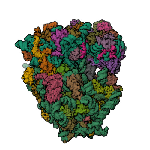
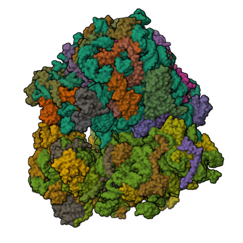
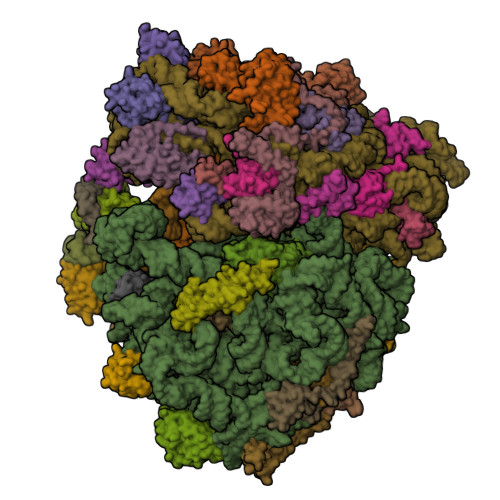
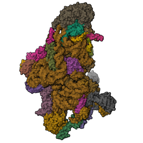
Cryo-EM structure of 3DVA component 1 of Escherichia coli que-PEC (paused elongation complex) RNA Polymerase plus preQ1 ligand
SM: PRF
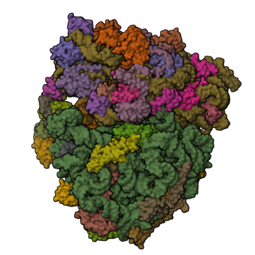
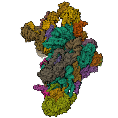
mRNA decoding in human is kinetically and structurally distinct from bacteria (Consensus LSU focused refined structure)
SM: 3H3 ANM
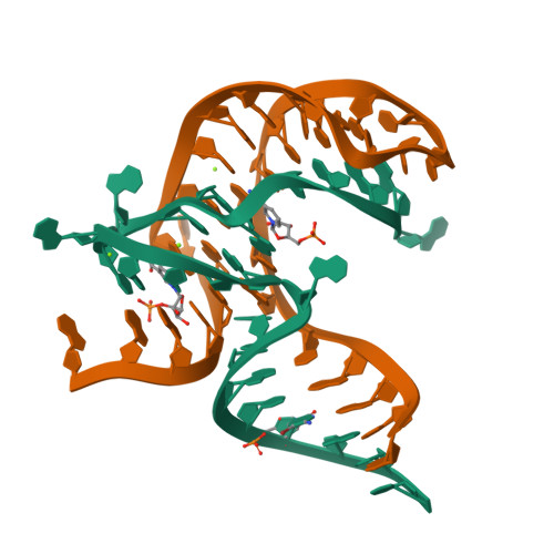
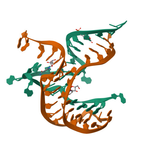
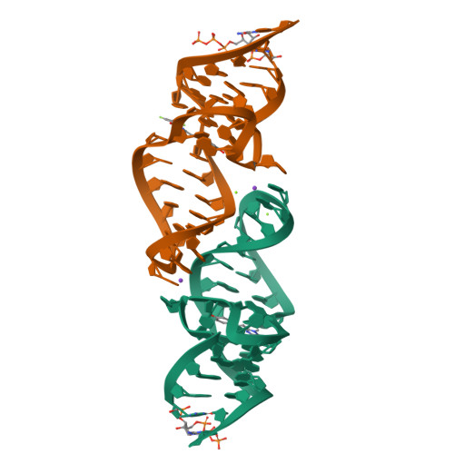
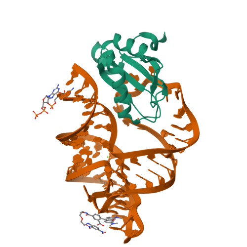
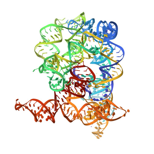
Crystal structure of NAD+ -II riboswitch in complex with NAD+
SM: NAD NAD
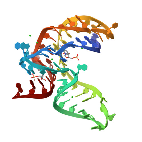
Crystal structure of NAD+ -II riboswitch in complex with NMN
SM: NMN NMN
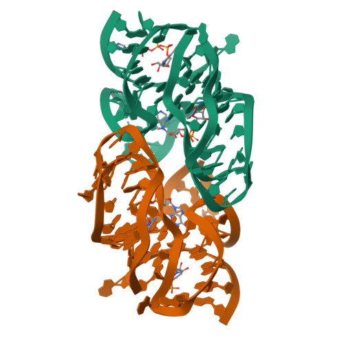
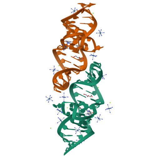
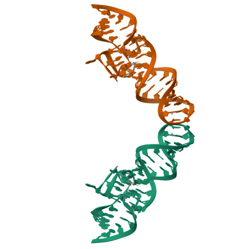
Crystal structure of NAD-II riboswitch (two strands) with NMN
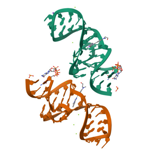
Crystal structure of NAD-II riboswitch (two strands) with NR
SM: NNR NNR
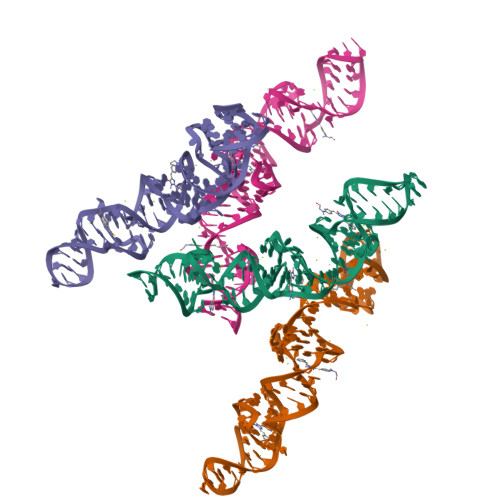
Crystal structure of NAD-II riboswitch (single strand) with NMN
Crystal structure of NAD-II riboswitch (single strand) with NAD
SM: NAD NAD NAD NMN
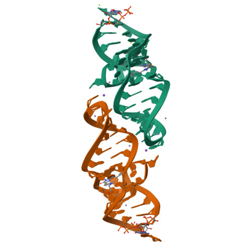
A new fluorescent RNA aptamer bound with N618
SM: O2I O2I
A new fluorescent RNA aptamer bound with N
SM: NI4 NI4
A new fluorescent RNA aptamer bound with N565
SM: NJL NJL
A new fluorescent RNA aptamer bound with N571
SM: O00 O00
A new fluorescent RNA aptamer bound with N, iridium hexammine soak
A new fluorescent RNA aptamer_III bound with N
SM: NI4 NI4 NI4 NI4 NI4 NI4
A new fluorescent RNA aptamer bound with N, manganese soak
Crystal structure of NAD-II riboswitch (two strands) with NMN at 1.67 angstrom
Interaction between a fluoroquinolone derivative KG022 and RNAs: effect of base pairs 3' adjacent to the bulge out residues
SM: 53D
The Tet-S1 state of G264A mutated Tetrahymena group I intron with 6nt 3'/5'-exon and 2-aminopurine nucleoside
SM: OJI
Dibekacin-bound E.coli 70S ribosome in the PURE system
SM: 84D 84D 84D 84D 84D 84D 84D 84D 84D 84D 84D 84D 84D SPM
Arbekacin-bound E.coli 70S ribosome in the PURE system
SM: 84G 84G 84G 84G 84G 84G 84G 84G 84G 84G SPM
Dibekacin-added human 80S ribosome
SM: 84D 84D 84D 84D 84D 84D 84D 84D 84D 84D 84D 84D 84D 84D 84D 84D 84D 84D 84D 84D 84D 84D 84D 84D
Arbekacin-added human 80S ribosome
SM: 84G 84G 84G 84G 84G 84G 84G 84G 84G 84G 84G 84G 84G 84G 84G 84G 84G 84G 84G 84G 84G 84G 84G
Crystal structure of RhoBAST complexed with TMR-DN
SM: V8C V8C V8C
Cryo-EM structure of the human 55S mitoribosome with Tigecycline
SM: T1C T1C T1C T1C
Cryo-EM structure of the human 39S mitoribosome with Tigecycline
SM: T1C
Cryo-EM structure of the human 80S ribosome with Tigecycline
SM: T1C T1C T1C T1C T1C T1C T1C T1C T1C
Crystal structure of Broccoli aptamer with DFHBI-1T
SM: 2ZY
Cryo-EM structure of the yeast 80S ribosome with tigecycline, Not5 and P-site tRNA
SM: T1C T1C T1C T1C T1C T1C
Crystal structure of Red Broccoli aptamer with OBI
SM: A1EBI A1EBI
Crystal structure of 2'-dG-III riboswitch with 2'-dG
SM: GNG
Crystal structure of 2'-dG-III riboswitch with guanosine
SM: GMP GMP
Crystal structure of the Candida albicans 80S ribosome in complex with Paromomycin (250uM)
SM: 3K5 3K5 PAR PAR PAR PAR PAR PAR PAR PAR
Structure of Candida albicans 80S ribosome in complex with mefloquine
SM: SPK YMZ YMZ
Crystal structure of the Candida albicans 80S ribosome in complex with Paromomycin (500umol)
SM: 3K5 3K5 PAR PAR PAR PAR PAR PAR PAR PAR PAR PAR PAR PAR PAR PAR PAR PAR PAR PAR PAR PAR PAR
Crystal structure of the Candida albicans 80S ribosome in complex with Paromomycin (2mM)
SM: 3K5 PAR PAR PAR PAR PAR PAR PAR PAR PAR PAR PAR PAR PAR PAR PAR PAR PAR PAR PAR PAR PAR PAR PAR PAR PAR PAR PAR PAR PAR PAR PAR PAR PAR PAR PAR PAR PAR PAR PAR PAR PAR PAR PAR PAR PAR PAR PAR PAR PAR PAR PAR PAR PAR PAR PAR
Crystal structure of the Candida albicans 80S ribosome in complex with geneticin G418 (rotated state)
SM: 3K5 3K5 GET GET
Structure of Oceanobacillus iheyensis group II intron in the presence of K+, Mg2+ and intronistat B
SM: EPE EPE SPM SPM VTE
Structure of Oceanobacillus iheyensis group II intron in the presence of K+, Mg2+ and ARN25850
SM: EPE EPE SPM VTR
Structure of Oceanobacillus iheyensis group II intron post first step of splicing in the presence of K+, Mg2+ and intronistat B
SM: EPE EPE EPE SPM SPM
Structure of Oceanobacillus iheyensis group II intron in the presence of K+, Mg2+, 5'-exon, and intronistat B after 2h30 soaking
SM: EPE SPM VTE
Structure of Oceanobacillus iheyensis group II intron in the presence of Na+, Mg2+ and intronistat B
SM: GDE
E167K RF2 on E. coli 70S release complex with UGG (Structure I)
SM: SPM
E167K RF2 on E. coli 70S release complex with UGG (Structure II)
E167K RF2 on E. coli 70S release complex with UGG (Structure III)
Ternary complex of translating ribosome, NAC and METAP1
SM: SPM SPM SPM
Mycoplasma pneumoniae 70S ribosome in chloramphenicol-treated cells
SM: CLM SPM SPM SPM SPM
Mycoplasma pneumoniae 70S ribosome with second S4 protein on large subunit
Mycoplasma pneumoniae large ribosomal subunit in chloramphenicol-treated cells
Escherichia coli paused disome complex (queueing 70S non-rotated closed PRE state)
SM: ATP ATP
E167K RF2 on E. coli 70S release complex with UAA
SM: SPM SPM
Crystal structure of RNA G2C4 repeats in complex with small synthetic molecule ANP77
SM: W53
mt-SSU assembly intermediate in GTPBP8 knock-out cells, state 1
SM: NAD SPM SRY
mt-SSU assembly intermediate in GTPBP8 knock-out cells, state 3
mt-SSU in GTPBP8 knock-out cells, state 4
Human 60S ribosomal subunit
SM: ATP SPM SPM SPM SPM SPM
Current selection range: to
Filter entries " Experimental resolution " between the two values. Global DB range: 0.6 – 11.2
Filter entries " Molecular weight (compound) " between the two values. Global DB range: 150 – 1000
Filter entries " Deposition date " between the two values. Global DB range: 1986 – 2027
Filter entries " Molecular weight (complex) " between the two values. Global DB range: 0 – 13090
Filter entries " Number of atoms " between the two values. Global DB range: 5 – 70
Filter entries " Number of interacting chains " between the two values. Global DB range: 1 – 4
Filter entries " Pocket size " between the two values. Global DB range: 15.9 – 394.0
Filter entries " SiteScore " between the two values. Global DB range: 0.3 – 1.3
Filter entries " Dscore " between the two values. Global DB range: -0.1 – 1.4
Filter entries " Exposure " between the two values. Global DB range: 0.1 – 1.0
Filter entries " Enclosure " between the two values. Global DB range: 0.3 – 1.0
Filter entries " Hydrophobicity " between the two values. Global DB range: 0.0 – 3.1
Filter entries " Hydrophilicity " between the two values. Global DB range: 0.2 – 2.8
Filter entries " Donor/Acceptor " between the two values. Global DB range: 0.0 – 5.0
Filter entries " Volume " between the two values. Global DB range: 21.2 – 1528.8