Harnessing RIBOnucleic acid - Small molecules Structures
Combine filters to refine your query. Each filter opens a dialog showing the values available in the data.
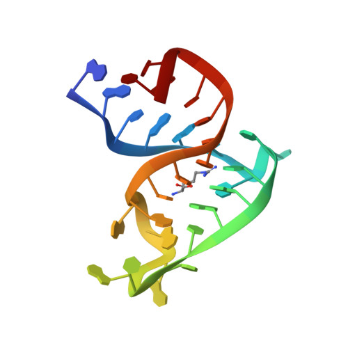
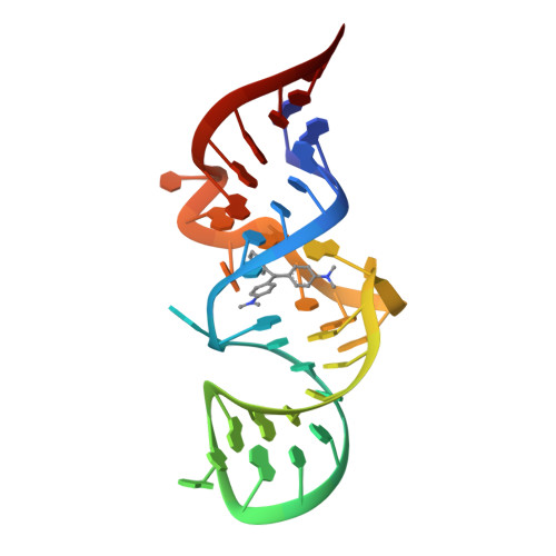
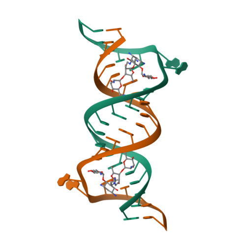
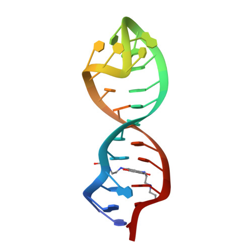
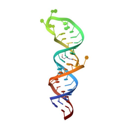
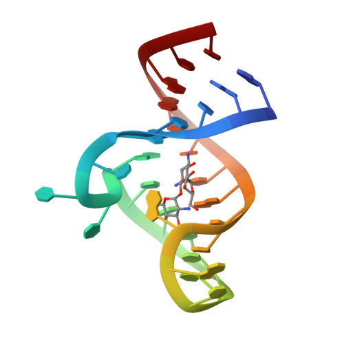
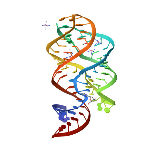
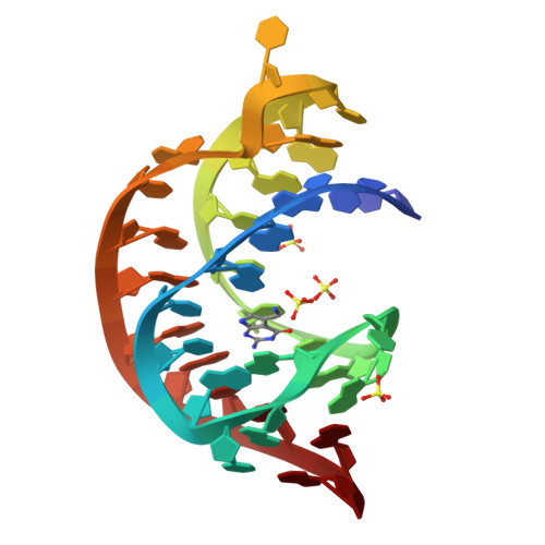
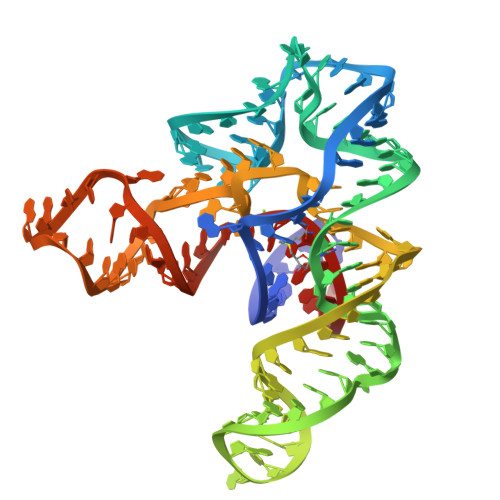
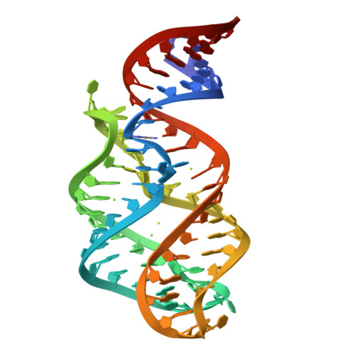
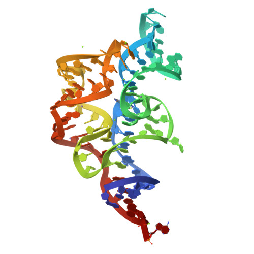
ARG-BOUND TAR RNA, NMR
SM: ARG
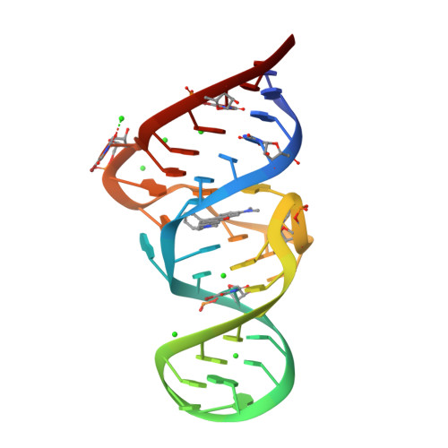
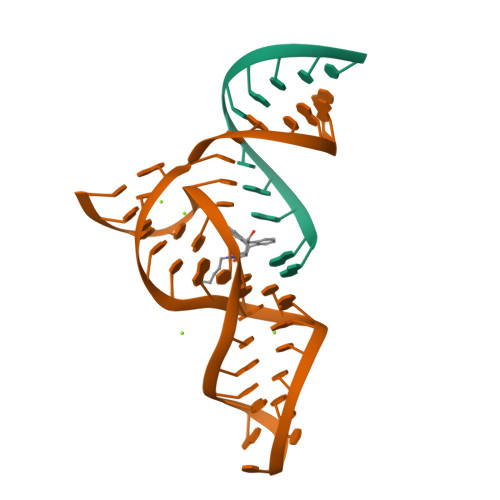
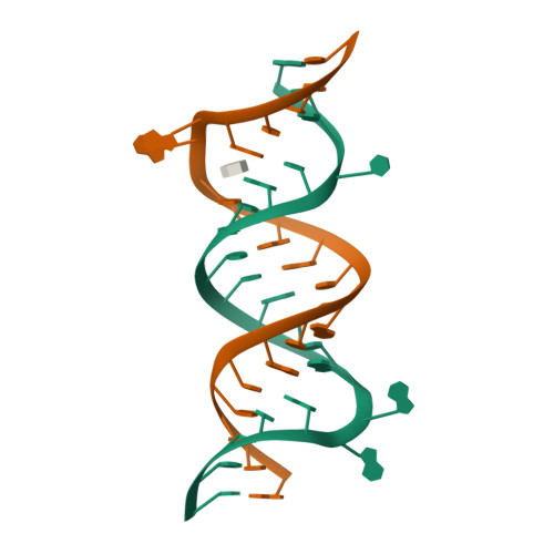
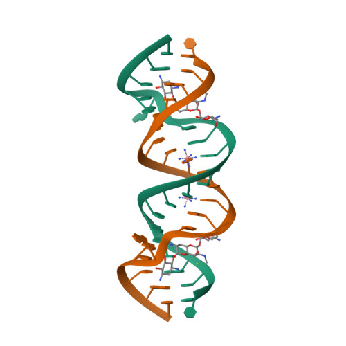
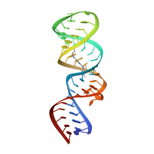
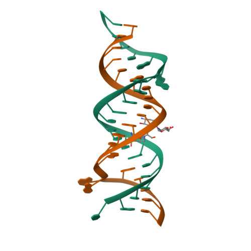
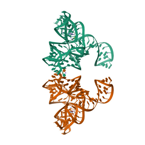
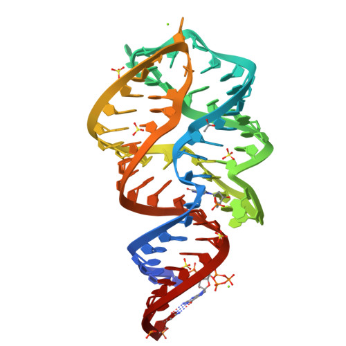
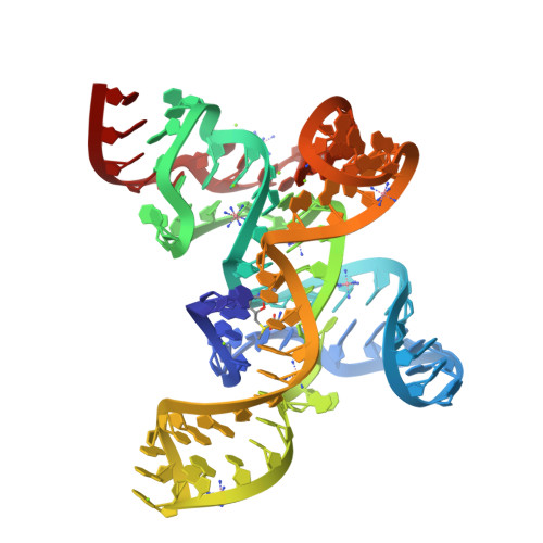
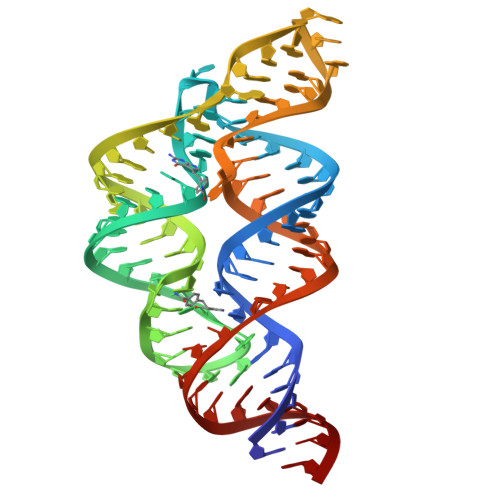
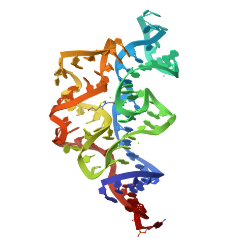
CRYSTAL STRUCTURE OF THE MALACHITE GREEN APTAMER COMPLEXED WITH TETRAMETHYL-ROSAMINE
SM: ROS
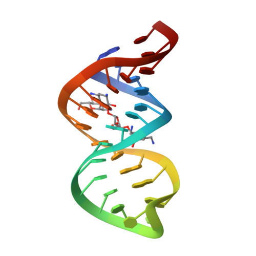
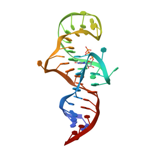
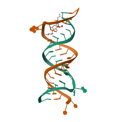
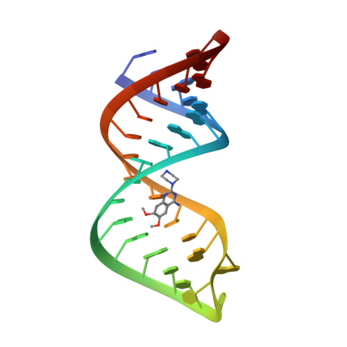
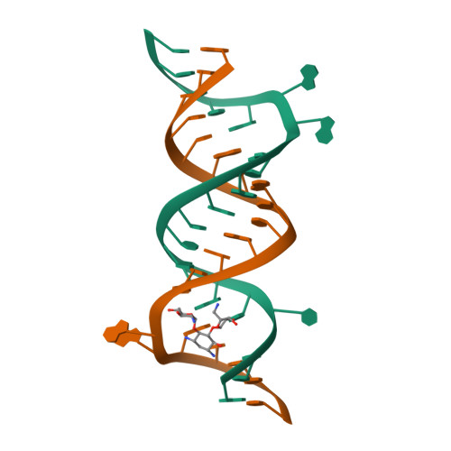
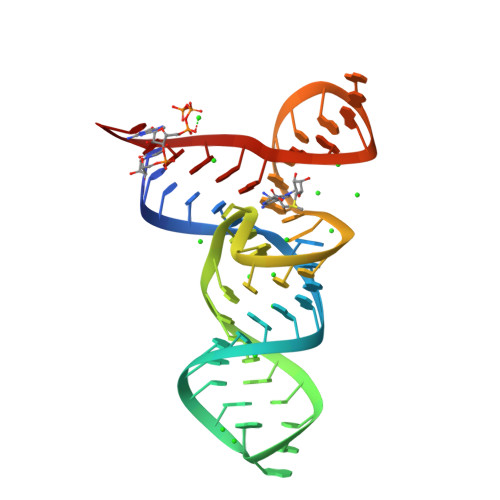
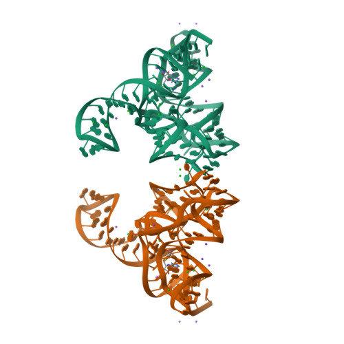
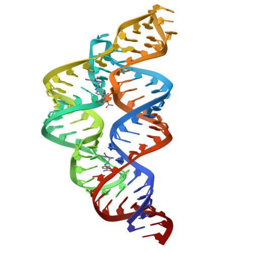
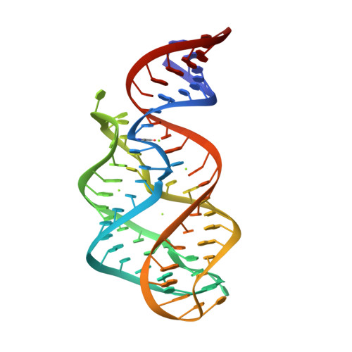
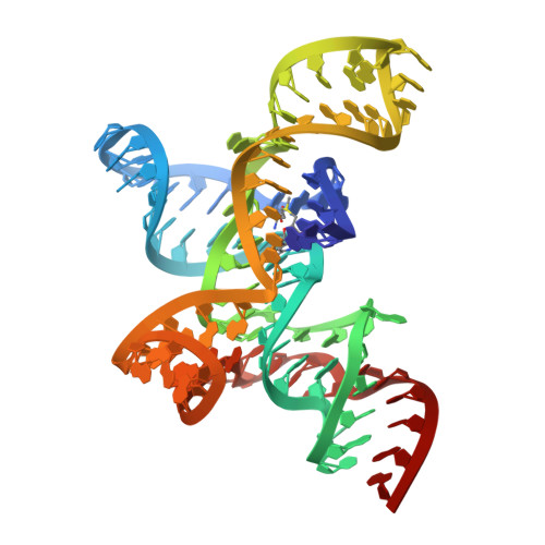
CRYSTAL STRUCTURE OF A BIOTIN-BINDING RNA PSEUDOKNOT
SM: BTN
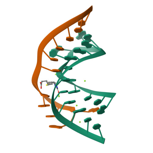
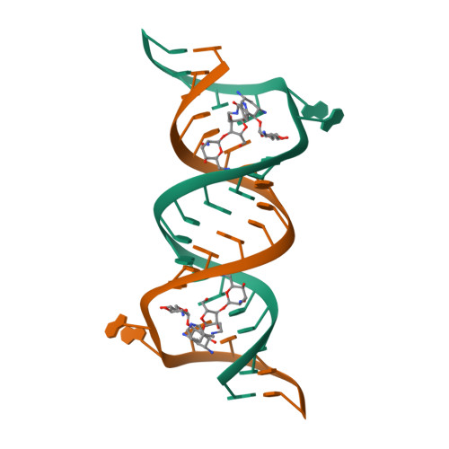
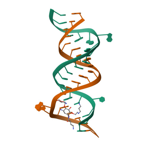
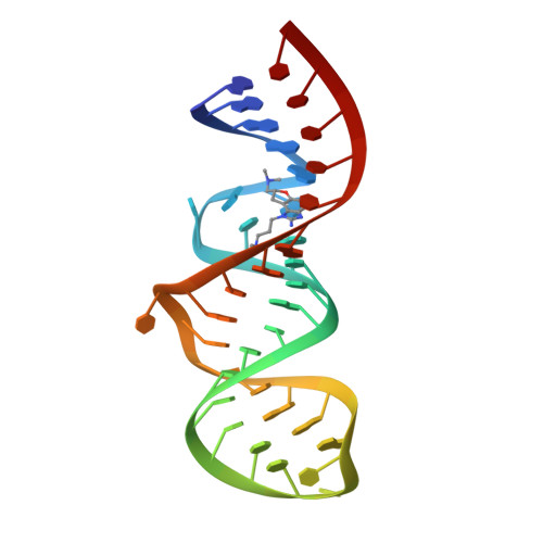
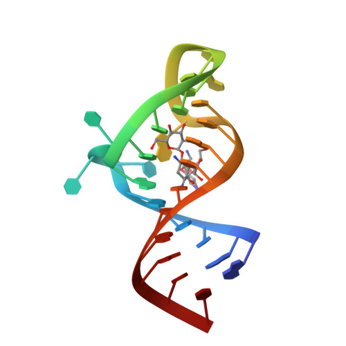
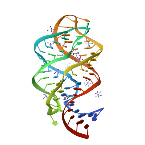
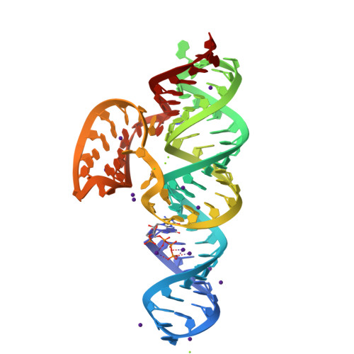
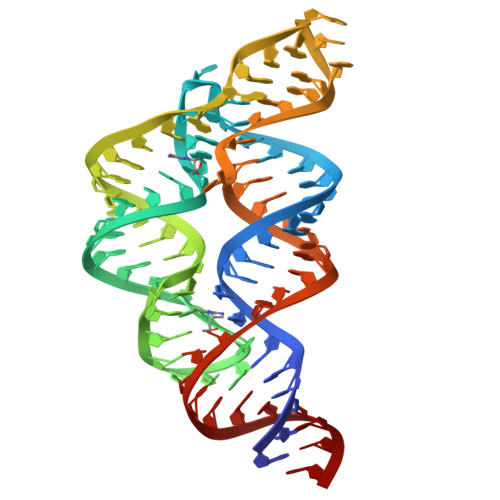
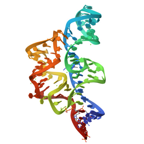
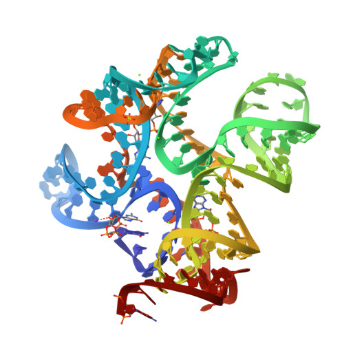
EUKARYOTIC DECODING REGION A-SITE RNA-PAROMOMYCIN COMPLEX
SM: PAR
Solution Structure of the Malachite Green RNA Binding Aptamer
SM: MGR
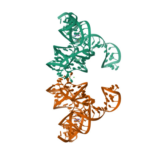
Crystal structure of the Diels-Alder ribozyme complexed with the product of the reaction between N-pentylmaleimide and covalently attached 9-hydroxymethylanthracene
SM: DAI DAI
Class I GTP aptamer
SM: GTP
Complex Between Paromomycin Derivative JS5-39 and the 16S-Rrna A-Site.
SM: JS5 JS5
Complex Between Paromomycin derivative JS4 and the 16S-Rrna A Site
SM: JS4 JS4
A-site RNA in complex with neamine
SM: XXX
Asite RNA + designer antibiotic
SM: AB9
SM: AB6
Crystal Structure of the Homo sapiens Cytoplasmic Ribosomal Decoding Site complexed with Apramycin
SM: AM2 AM2
Structural basis for stabilization of the tau pre-mRNA splicing regulatory element by Novantrone (Mitoxantrone)
SM: MIX
Inhibitor Induced Structural Change in the HCV IRES Domain IIa RNA
SM: ISH
SM: ISI
NMR structure of stem-loop 4 from the human 7SK snRNA in complex with arginine
Structure of the HIV-1 frameshift site RNA bound to a small molecule inhibitor of viral replication
SM: L94
Solution structure of small molecule-influenza RNA complex
SM: 0EC
Solution NMR-structure of the neomycin sensing riboswitch RNA bound to paromomycin
Solution NMR Structure of the 27 nucleotide engineered neomycin sensing riboswitch RNA-ribostamycin complex
SM: RIO
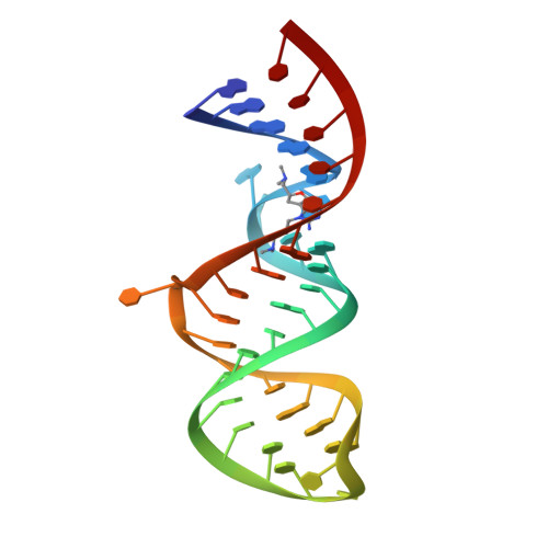
Crystal Structure of the Homo sapiens Cytoplasmic Ribosomal Decoding Site in presence of paromomycin
Crystal Structure of the Prokaryotic Ribosomal Decoding Site Complexed with Paromamine Derivative NB30
SM: N30
Crystal structure of the SAM-I riboswitch A94G U34 G18U G19U variant in complex with SAM
SM: SAM
SAM-I riboswitch with a G2nA mutation in the Kink turn in complex with S-adenosylmethionine
Structure of an RNA-2'-deoxyguanosine complex
SM: GNG GNG
Crystal Structure of the SMK box (SAM-III) Riboswitch with SAM
Crystal structure of guanine riboswitch C74U mutant bound to 6-chloroguanine
SM: 6GU
Crystal structure of guanine riboswitch bound to 6-O-methylguanine
SM: 6GO
Cocrystal structure of a class-I preQ1 riboswitch
SM: PRF PRF PRF
Guanine riboswitch bound to 6-chloroguanine
Crystal structure of the guanine riboswitch C74U mutant bound to 6-O-methylguanine
Guanine riboswitch A21G,U75C mutant bound to 6-chloroguanine
Crystal Structure of the G20A mutant c-di-GMP riboswith bound to c-di-GMP
SM: C2E
Crystal Structure of the C92U mutant c-di-GMP riboswith bound to c-di-GMP
Native structure of a c-di-GMP riboswitch from V. cholerae
Structure of the s-adenosylhomocysteine riboswitch at 3.0A
SM: SAH
Structure of a c-di-GMP-II riboswitch from C. acetobutylicum bound to c-di-GMP
SM: C2E C2E C2E C2E
Structural analysis of a class I PreQ1 riboswitch aptamer in the metabolite-bound state
SM: PRF
Crystal structure of the 2'- Deoxyguanosine riboswitch bound to 2'-deoxyguanosine-5'-monophosphate
SM: DGP DGP
SAM-I riboswitch containing the T. solenopsae Kt-23 in complex with S- adenosyl methionine
Structure of a class II preQ1 riboswitch reveals ligand recognition by a new fold
Bacillus subtilis yitJ S box/SAM-I riboswitch
Structure of the SAM-I/IV riboswitch (env87(deltaU92, deltaG93))
Structure of the THF riboswitch
SM: FFO FFO
Structure of the THF riboswitch bound to tetrahydrobiopterin
SM: H4B H4B
Structure of the THF riboswitch bound to pemetrexed
SM: LYA LYA
X-ray crystal structure of the M6" riboswitch aptamer bound to pyrimido[4,5-d]pyrimidine-2,4-diamine (PPDA)
SM: 29G
X-ray crystal structure of the M6C" riboswitch aptamer bound to 2-aminopyrimido[4,5-d]pyrimidin-4(3H)-one (PPAO)
SM: 29H
Crystal structure of the E. coli thiM riboswitch in complex with 5-(azidomethyl)-2-methylpyrimidin-4-amine
SM: 2QB 2QB
Crystal structure of the E. coli thiM riboswitch in complex with (4-(1,2,3-thiadiazol-4-yl)phenyl)methanamine
SM: 2QC
Crystal structure of the E. coli thiM riboswitch in complex with thiamine
SM: VIB
Structure of the SAM-I/IV riboswitch (env87(deltaU92))
Thermoanaerobacter pseudethanolicus c-di-AMP riboswitch
SM: 2BA 2BA
Thermovirga lienii c-di-AMP riboswitch
ydao riboswitch binding to c-di-AMP
structure of ydao riboswitch binding with c-di-dAMP
Crystal Structure of a PreQ1 Riboswitch
Crystal structure of the Spinach RNA aptamer in complex with DFHBI, barium ions
SM: 38E
Crystal structure of the Spinach RNA aptamer in complex with DFHBI, magnesium ions
Current selection range: to
Filter entries " Experimental resolution " between the two values. Global DB range: 0.6 – 11.2
Filter entries " Molecular weight (compound) " between the two values. Global DB range: 150 – 1000
Filter entries " Deposition date " between the two values. Global DB range: 1986 – 2027
Filter entries " Molecular weight (complex) " between the two values. Global DB range: 0 – 13090
Filter entries " Number of atoms " between the two values. Global DB range: 5 – 70
Filter entries " Number of interacting chains " between the two values. Global DB range: 1 – 4
Filter entries " Pocket size " between the two values. Global DB range: 15.9 – 394.0
Filter entries " SiteScore " between the two values. Global DB range: 0.3 – 1.3
Filter entries " Dscore " between the two values. Global DB range: -0.1 – 1.4
Filter entries " Exposure " between the two values. Global DB range: 0.1 – 1.0
Filter entries " Enclosure " between the two values. Global DB range: 0.3 – 1.0
Filter entries " Hydrophobicity " between the two values. Global DB range: 0.0 – 3.1
Filter entries " Hydrophilicity " between the two values. Global DB range: 0.2 – 2.8
Filter entries " Donor/Acceptor " between the two values. Global DB range: 0.0 – 5.0
Filter entries " Volume " between the two values. Global DB range: 21.2 – 1528.8