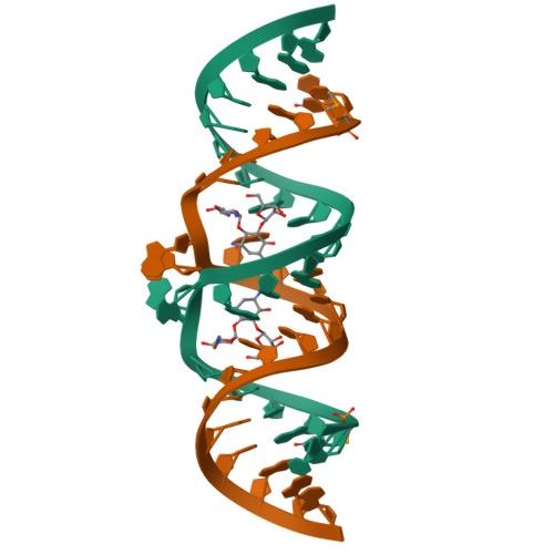
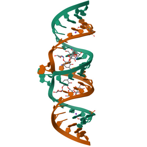
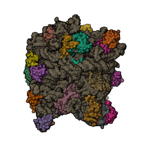
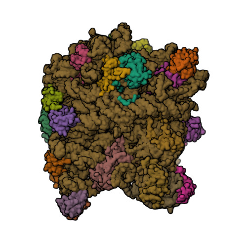
Harnessing RIBOnucleic acid - Small molecules Structures
Combine filters to refine your query. Each filter opens a dialog showing the values available in the data.
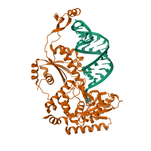
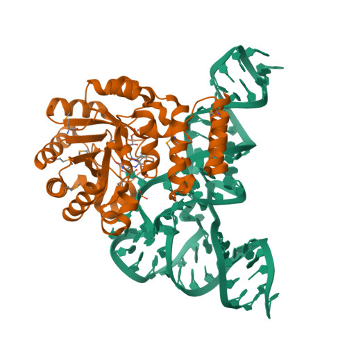
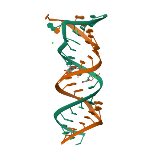
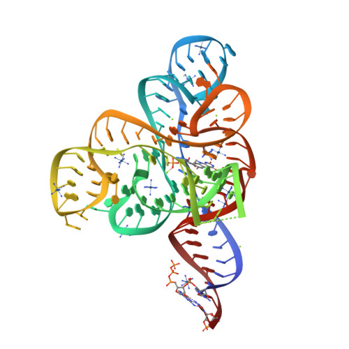
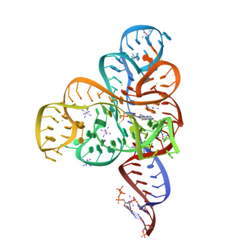
Complex structure of AFCCA with tRNAminiDCU and ATP
SM: ATP
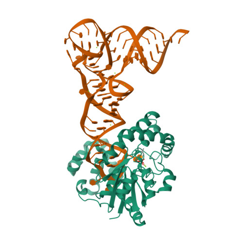
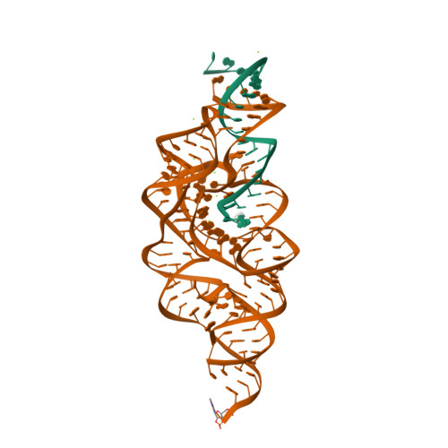
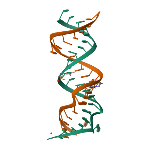
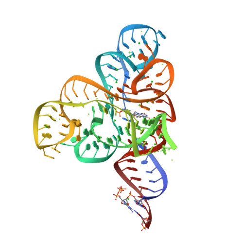
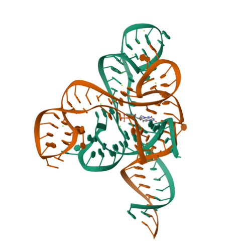
Complex structure of AFCCA with tRNAminiDU and CTP
SM: CTP
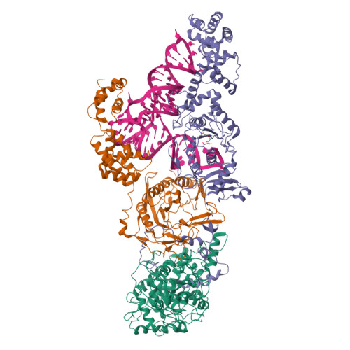
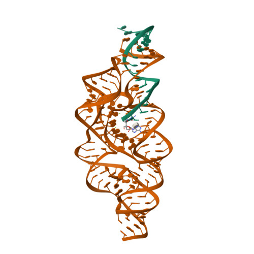
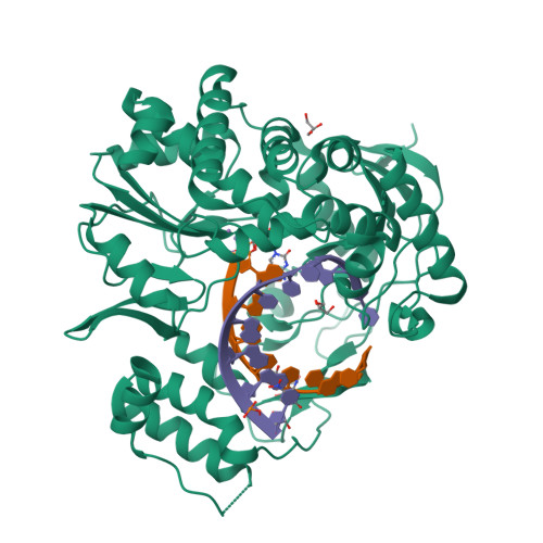
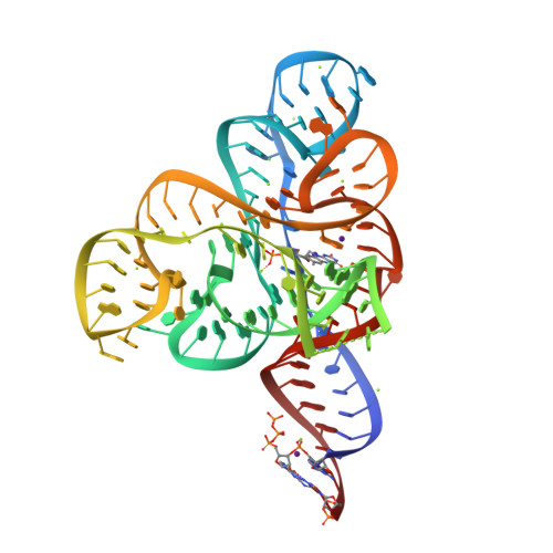
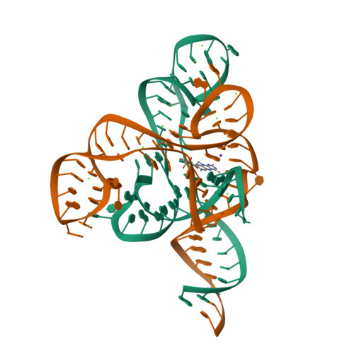
Crystal structure of tRNA modification enzyme MiaA in the complex with tRNA(Phe) and DMASPP
SM: DST
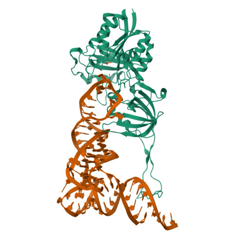
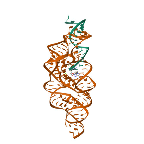
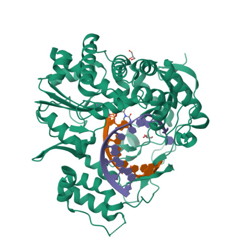
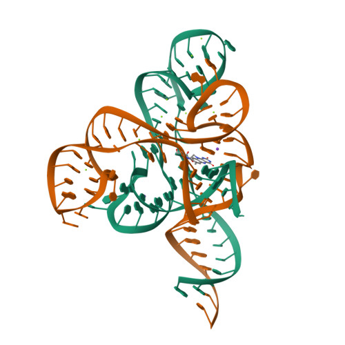
Cryo-EM structure of the yeast RNA polymerase II elongation complex with 19-mer RNA in State III (TL-open), in the presence of substrate ATP
Cryo-EM structure of the yeast RNA polymerase II elongation complex with 19-mer RNA in State V (TL-closed), in the presence of substrate ATP
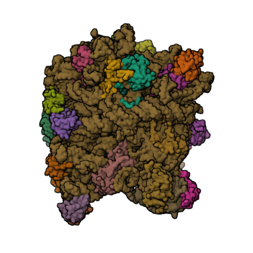
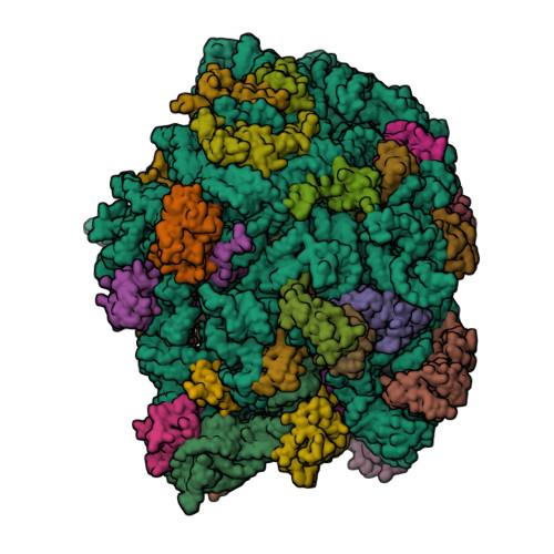
Cryo-EM structure of SKM-M.smegmatis 70S ribosomal complex
SM: A1JAI
RNA-L-G monomer complex
SM: 0G 0G 0G 0G
RNA-dGMP complex with L-G-terminal primer
SM: DGP DGP DGP DGP DGP DGP DGP DGP
RNA-dGMP complex
SM: DGP DGP DGP DGP
Crystal structure of Thermotoga maritima nondiscriminating glutamyl-tRNA synthetase in complex with tRNAGln and a glutamyl-AMP analog
SM: GSU GSU GSU
Crystal structure of the glutamine transamidosome from Thermotoga maritima in the glutamylation state.
SM: GSU
Crystal structure of the TiaS-tRNA(Ile2)-AMPCPP-agmatine complex
SM: APC
Structure of viral RNA polymerase complex 1
SM: GH3 GH3
Structure of viral RNA polymerase complex 4
SM: GH3
Structure of viral RNA polymerase complex 5
Structure of viral RNA polymerase complex 6
SM: CH1
tRNA-dihydrouridine synthase from Thermus thermophilus in complex with tRNA fragment
SM: FMN FMN
tRNA-dihydrouridine synthase from Thermus thermophilus in complex with tRNA
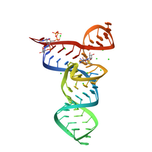
T. tengcongensis glmS ribozyme with G40A mutation, bound to glucosamine-6-phosphate
SM: GLP
T. tengcongensis glmS ribozyme with G40A mutation, bound to glucosamine-6-phosphate and a substrate RNA with a 2'5'-phosphodiester linkage
T. tengcongensis glmS ribozyme bound to glucosamine-6-phosphate and a substrate RNA with a 2'5'-phosphodiester linkage
Crystal Structure of the Homo sapiens Mitochondrial Ribosomal Decoding Site in the Presence of SrCl2 (A1555G mutant, Br-derivative)
SM: PAR
Crystal Structure of the Homo sapiens Mitochondrial Ribosomal Decoding Site in the presence of nonspecifically bound paromomycin (A1555G mutant, Br-derivative)
Norwalk Virus polymerase bound to 5-nitrocytidine triphosphate and primer-template RNA
SM: N5C
Norwalk Virus polymerase bound to cytidine 5'-triphosphate and primer-template RNA
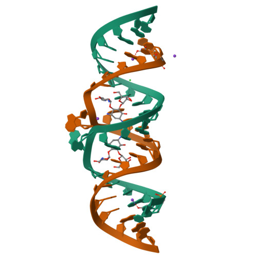
Crystal structure of HIV-1 subtype F DIS extended duplex RNA bound to ribostamycin
SM: RIO RIO
Crystal structure of HIV-1 subtype F DIS extended duplex RNA bound to paromomycin
SM: PAR PAR
Crystal structure of HIV-1 subtype F DIS extended duplex RNA bound to lividomycin
SM: LIV LIV
Crystal Structure of HIV-1 subtype F DIS extended duplex RNA bound to neomycin
SM: NMY NMY
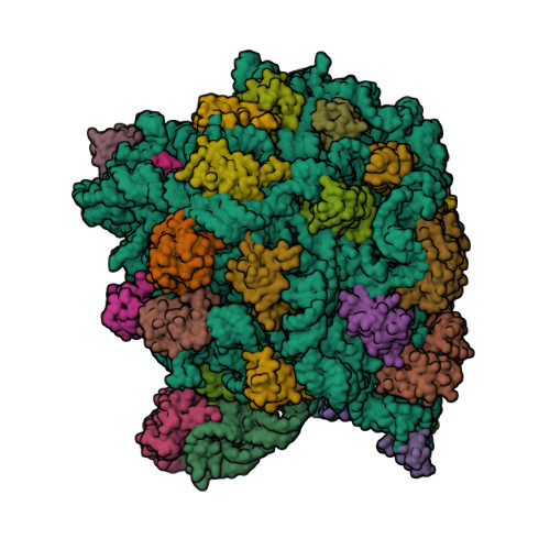
Co-crystal Structure of Anisomycin Bound to the 50S Ribosomal Subunit
SM: ANM
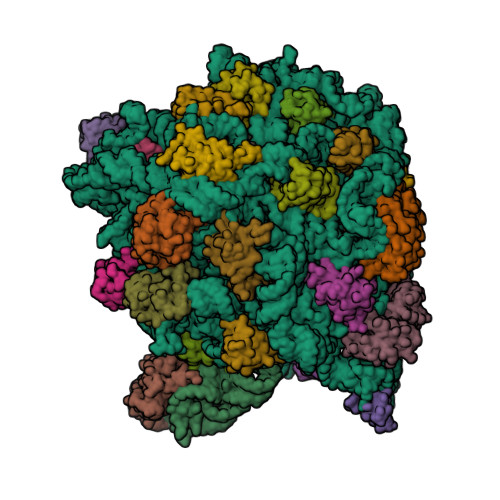
The structure of CCA and CCA-Phe-Cap-Bio bound to the large ribosomal subunit of Haloarcula marismortui
SM: PHE
The Structure of CA and CCA-PHE-CAP-BIO Bound to the Large Ribosomal Subunit of Haloarcula Marismortui
The structure of the antibiotic LINEZOLID bound to the large ribosomal subunit of HALOARCULA MARISMORTUI
SM: ZLD
The structure of an enhanced oxazolidinone inhibitor bound to the 50S ribosomal subunit of H. marismortui
SM: SLD
Structural basis of thiamine pyrophosphate analogues binding to the eukaryotic riboswitch
SM: TPP TPP
Structure of the eukaryotic TPP-specific riboswitch bound to the antibacterial compound pyrithiamine pyrophosphate
SM: PYI PYI
Structure of the thiamine pyrophosphate-specific riboswitch bound to oxythiamine pyrophosphate
SM: D2X D2X
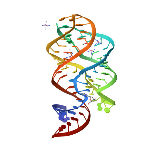
CRYSTAL STRUCTURE OF THE THERMOTOGA MARITIMA LYSINE RIBOSWITCH BOUND TO S-(2-aminoethyl)-L-cysteine
SM: SLZ
Crystal structure of the Thermotoga maritima lysine riboswitch bound to lysine
SM: 1PE 1PE 1PE
Crystallization of the Thermotoga maritima lysine riboswitch bound to lysine, Cs+ Soak
SM: 1PE
Crystallization of the Thermotoga maritima lysine riboswitch bound to lysine, IRIDIUM HEXAMINE SOAK
Crystallization of the Thermotoga maritima lysine riboswitch bound to homoarginine
SM: 1PE HRG
Crystallization of the Thermotoga maritima lysine riboswitch bound to N6-1-iminoethyl-L-Lysine
SM: IEL
The oxazolidinone antibiotics perturb the ribosomal peptidyl-transferase center and effect tRNA positioning
Structure of an RNA-2'-deoxyguanosine complex
SM: GNG GNG
Crystal structure of HIV-1 subtype F DIS extended duplex RNA bound to ribostamycin (U267OMe)
Crystal Structure of the SMK box (SAM-III) Riboswitch with SAM
SM: SAM
Crystal Structures of the SMK box (SAM-III) Riboswitch with SAH
SM: SAH
Crystal Structures of the SMK box (SAM-III) Riboswitch with Se-SAM
SM: EEM
Crystal structure of an in vitro evolved tetracycline aptamer and artificial riboswitch
SM: CTC
Crystallographic snapshots of eukaryotic dimethylallyltransferase acting on tRNA: Insight into tRNA recognition and reaction mechanism
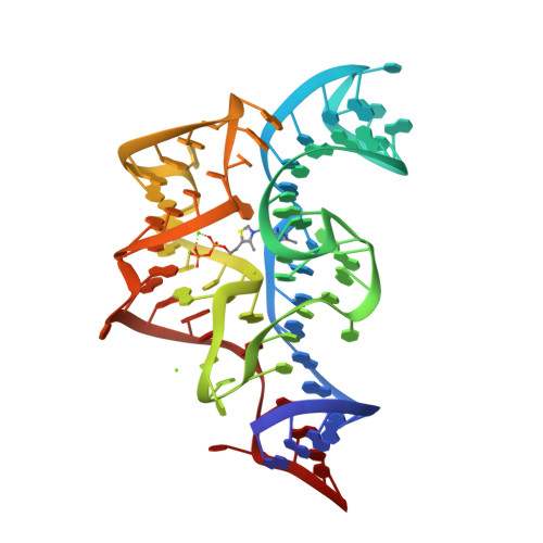
Crystal structure of the FMN riboswitch bound to FMN
SM: FMN
Crystal structure of the FMN riboswitch bound to FMN, iridium hexamine soak.
Crystal structure of the FMn riboswitch bound to FMN, Ba2+ soak.
Crystal structure of the FMN riboswitch bound to FMN, Cs+ soak.
Crystal structure of the FMN riboswitch bound to FMN, Mn2+ soak.
Crystal structure of the FMN riboswitch bound to FMN, cobalt hexammine soak.
Crystal structure of the FMN riboswitch bound to FMN, split RNA.
Crystal structure of the FMN riboswitch bound to riboflavin.
SM: RBF
Crystal structure of the FMN riboswitch bound to roseoflavin
SM: RS3
Current selection range: to
Filter entries " Experimental resolution " between the two values. Global DB range: 0.6 – 11.2
Filter entries " Molecular weight (compound) " between the two values. Global DB range: 150 – 1000
Filter entries " Deposition date " between the two values. Global DB range: 1986 – 2027
Filter entries " Molecular weight (complex) " between the two values. Global DB range: 0 – 13090
Filter entries " Number of atoms " between the two values. Global DB range: 5 – 70
Filter entries " Number of interacting chains " between the two values. Global DB range: 1 – 4
Filter entries " Pocket size " between the two values. Global DB range: 15.9 – 394.0
Filter entries " SiteScore " between the two values. Global DB range: 0.3 – 1.3
Filter entries " Dscore " between the two values. Global DB range: -0.1 – 1.4
Filter entries " Exposure " between the two values. Global DB range: 0.1 – 1.0
Filter entries " Enclosure " between the two values. Global DB range: 0.3 – 1.0
Filter entries " Hydrophobicity " between the two values. Global DB range: 0.0 – 3.1
Filter entries " Hydrophilicity " between the two values. Global DB range: 0.2 – 2.8
Filter entries " Donor/Acceptor " between the two values. Global DB range: 0.0 – 5.0
Filter entries " Volume " between the two values. Global DB range: 21.2 – 1528.8