Harnessing RIBOnucleic acid - Small molecules Structures
Combine filters to refine your query. Each filter opens a dialog showing the values available in the data.
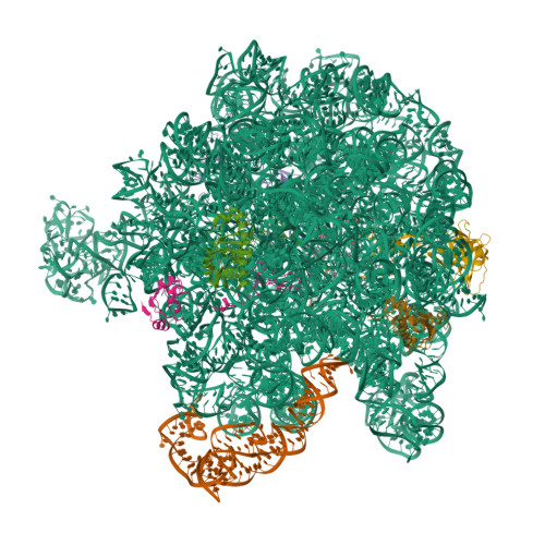
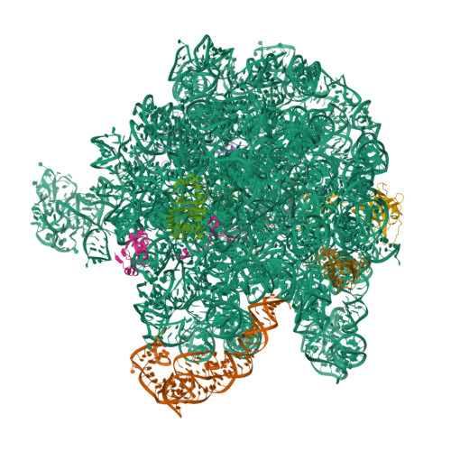
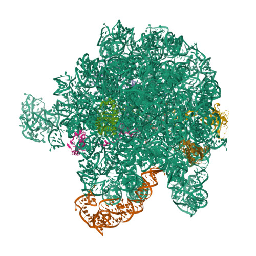
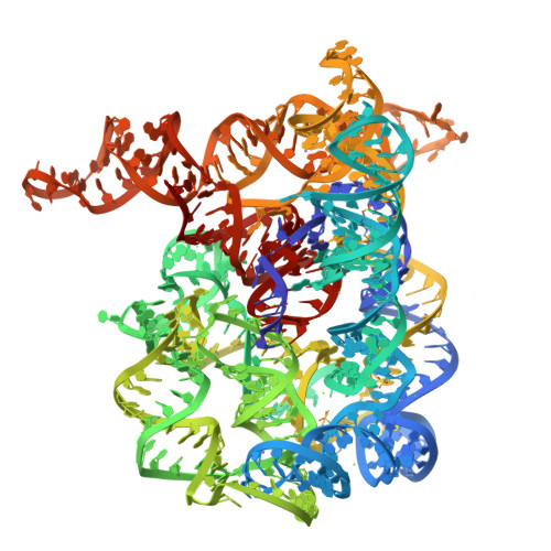
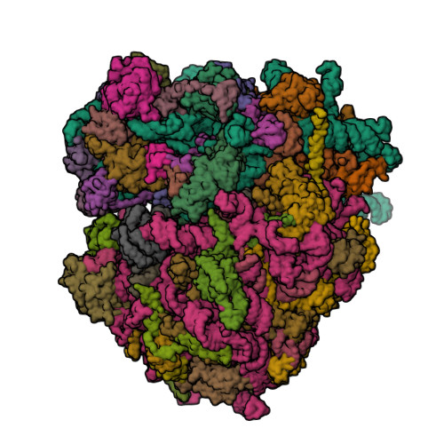
E. coli 50S ribosome bound to compounds 46 and VS1
SM: O7V
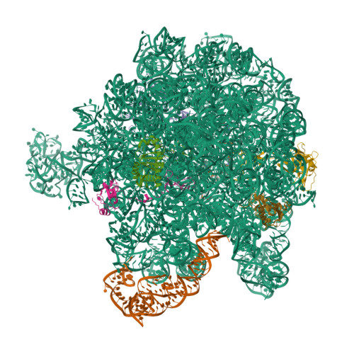
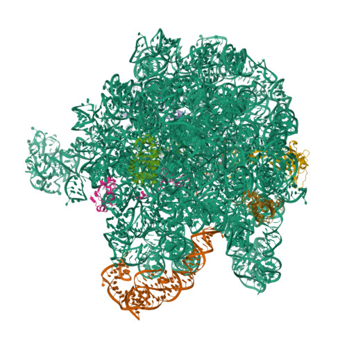
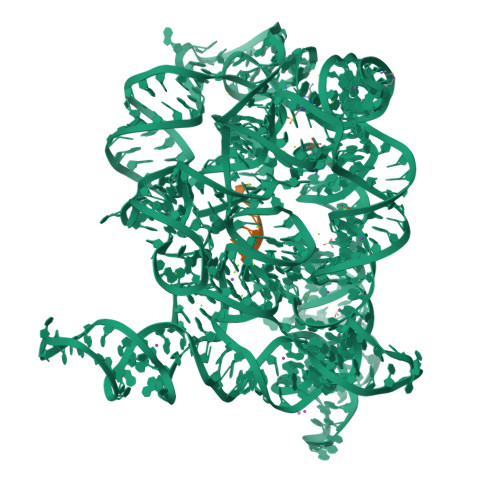
E. coli 50S ribosome bound to compound 47
SM: O7S
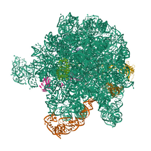
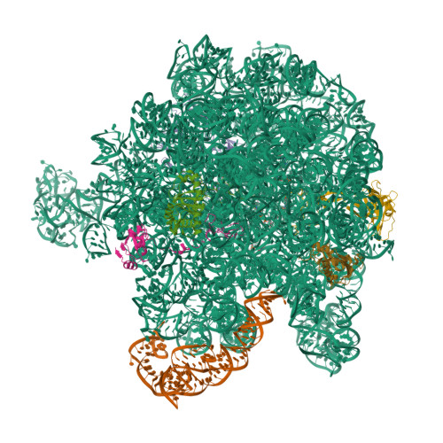
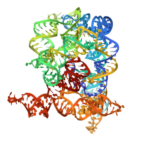
E. coli 50S ribosome bound to compound 46
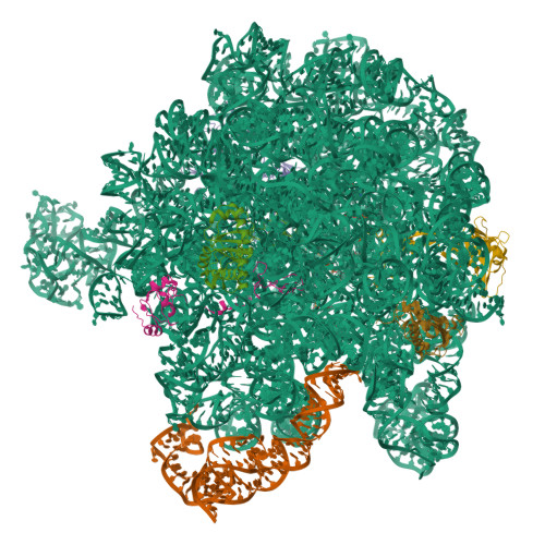
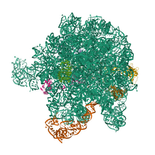
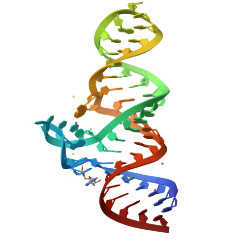
E. coli 50S ribosome bound to compound 40q
SM: O7Y
E. coli 50S ribosome bound to compound 21
SM: O8D
E. coli 50S ribosome bound to VM2
SM: O8J
E. coli 50S ribosome bound to compound 40o
SM: O8P
E. coli 50S ribosome bound to compound 40e
SM: O8S
E. coli 50S ribosome bound to compound 41q
SM: O8V
Structure of the iMango-III fluorescent aptamer at room temperature.
SM: OXV
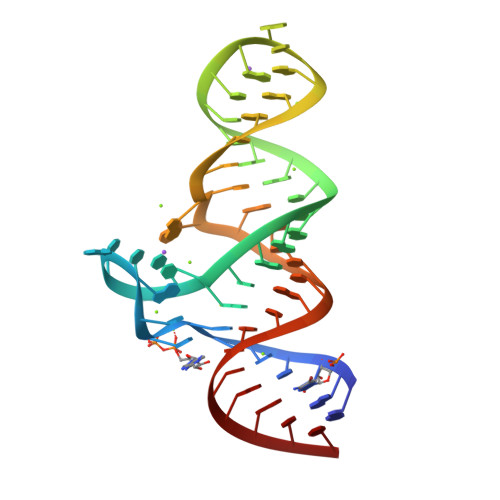
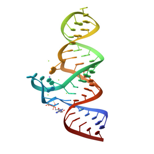
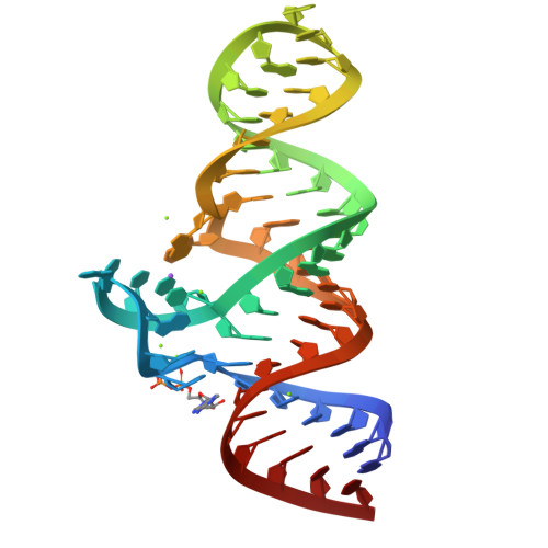
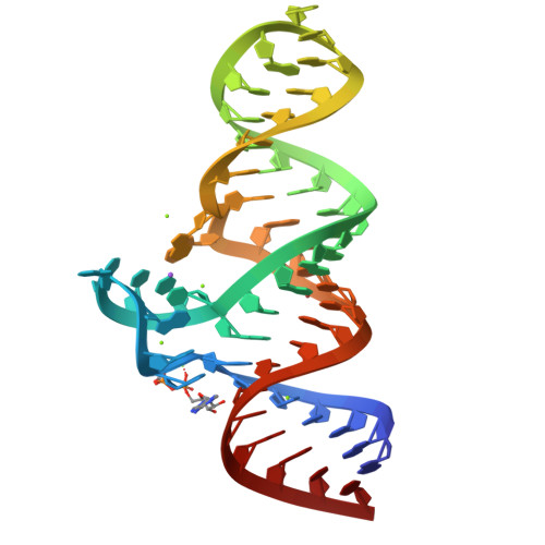
X-ray crystal structure of the tetrahydrofolate riboswitch aptamer bound to 5-deazatetrahydropterin
SM: T0A T0A
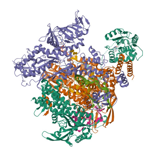
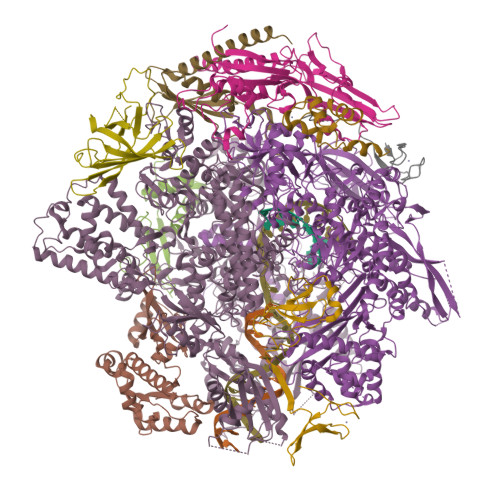
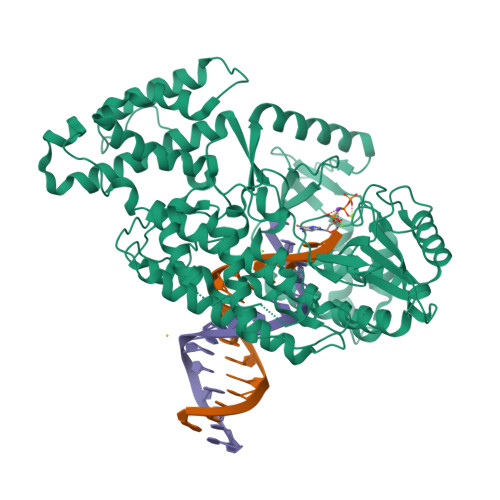
Crystal structure of influenza B polymerase initiation state with capped 14-mer RNA primer and CTP
SM: CTP
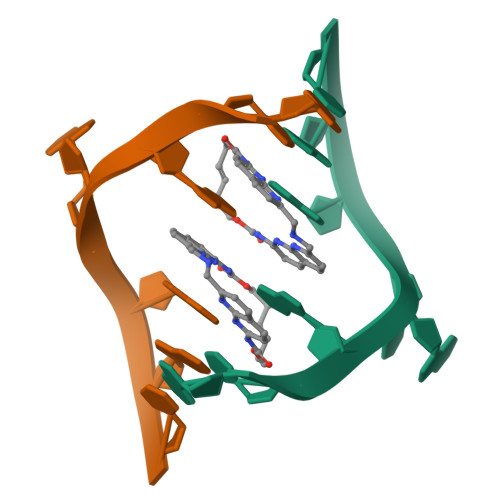
Crystal structure of seleno-derivative CAG repeats with synthetic CMBL3a compound
SM: J48 J48
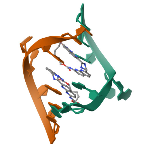
Crystal structure of CAG repeats with synthetic CMBL3a compound (model I)
SM: J48 J48 J48 J48
Crystal structure of CAG repeats with synthetic CMBL3b compound
Crystal structure of seleno-derivative CAG repeats with synthetic CMBL4 compound
SM: J4H
Structure of a bacterial 50S ribosomal subunit in complex with the novel quinoxolidinone antibiotic cadazolid
SM: AMP JJH
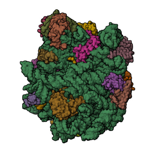
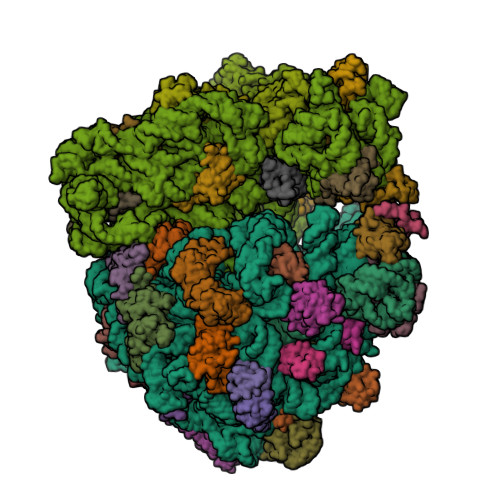
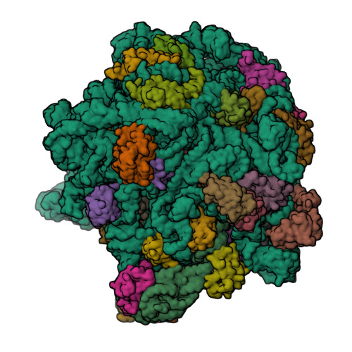
High-resolution cryo-EM structure of the human 80S ribosome
SM: HMT HYG
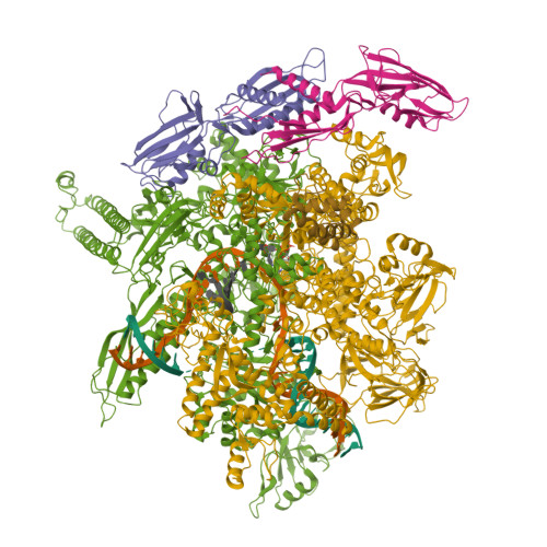
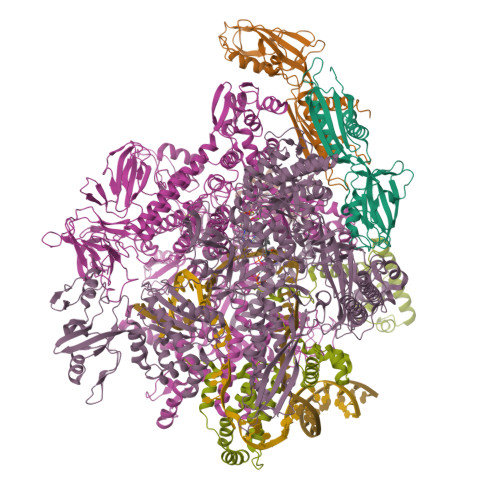
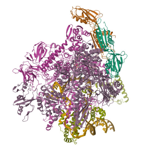
Cryo-EM structure of E. coli RNA polymerase elongation complex bound to CTP substrate
Structure of human mitochondrial 28S ribosome in complex with mitochondrial IF3
SM: NAD SPM SRY
Structure of human mitochondrial 28S ribosome in complex with mitochondrial IF2 and IF3
Erythromycin Resistant Staphylococcus aureus 70S ribosome (delta R88 A89 uL22) in complex with erythromycin.
SM: ERY
Erythromycin Resistant Staphylococcus aureus 50S ribosome (delta R88 A89 uL22) in complex with erythromycin.
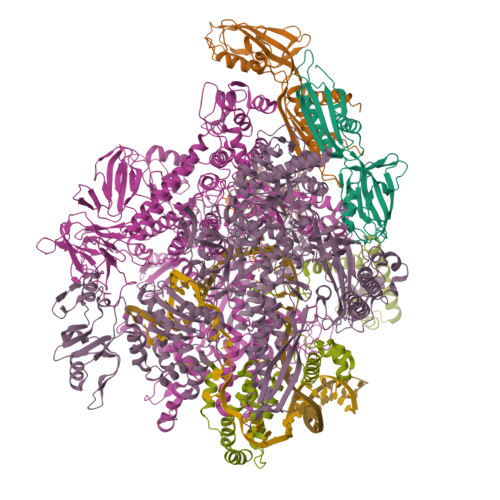
Bat Influenza A polymerase elongation complex with incoming UTP analogue (core + endonuclease only)
SM: 2KH
Bat Influenza A polymerase elongation complex with incoming UTP analogue (complete polymerase)
Structure of Oceanobacillus iheyensis group II intron G-mutant (C289G/C358G/G385C) in the presence of K+, Mg2+ and 5'-exon
SM: EPE EPE EPE SPM SPM
Structure of Oceanobacillus iheyensis group II intron G-mutant (C289G/C358G/G385C) in the presence of Na+, Mg2+ and 5'-exon
SM: EPE EPE EPE
Structure of Oceanobacillus iheyensis group II intron U-mutant (C289U/C358U/G385A) in the presence of K+, Mg2+ and 5'-exon
SM: EPE EPE SPM SPM
Structure of Oceanobacillus iheyensis group II intron U-mutant (C289U/C358U/G385A) in the presence of Na+, Mg2+ and 5'-exon
Crystal structure of the ADP-binding domain of the NAD+ riboswitch with Adenosine monophosphate (AMP)
SM: AMP
Crystal structure of the ADP-binding domain of the NAD+ riboswitch with Nicotinamide adenine dinucleotide, reduced (NADH)
SM: NAI
Crystal structure of the ADP-binding domain of the NAD+ riboswitch with Adenosine diphosphate (ADP)
SM: ADP
Crystal structure of the ADP-binding domain of the NAD+ riboswitch with Adenosine 5-triphosphate (ATP)
SM: ATP
Crystal structure of the ADP-binding domain of the NAD+ riboswitch with Cordycepin 5-triphosphate (3-dATP)
SM: 3AT
Crystal structure of the ADP-binding domain of the NAD+ riboswitch with N6-Methyl-adenosine-5'-triphosphate (m6ATP)
SM: N6E
Crystal structure of the ADP-binding domain of the NAD+ riboswitch with Nicotinamide adenine dinucleotide (NAD+)
SM: NAD
Crystal structure of the ADP-binding domain of the NAD+ riboswitch with Adenosine 3-phosphate 5-phosphosulfate (APPS)
SM: PPS
Crystal structure of the ADP-binding domain of the NAD+ riboswitch with Nicotinamide adenine dinucleotide, reduced (NADH); soaking with Manganese(II) (Mn2+)
Yeast 80S ribosome in complex with eIF5A and decoding A-site and P-site tRNAs.
SM: 3HE
In situ structure of BmCPV RNA-dependent RNA polymerase at elongation state
SM: ATP UTP
RNA-monomer complex containing pyrophosphate linkage
SM: EQ4 EQ4
RNA hairpin structure containing one TNA nucleotide as template
SM: C5P
RNA hairpin structure containing one TNA nucleotide as primer
RNA duplex, bound with TNA monomer
SM: TG TG
RNA hairpin, bound with TNA monomer
SM: TG
RNA duplex bound with TNA 3'-3' imidazolium dimer
SM: Q1V Q1V
Structure of guanine riboswitch bound to N2-acetyl guanine
SM: Q44
Guanine riboswitch bound to 8-aminoguanine
SM: ANG
Guanine riboswitch bound to O6-cyclohexylmethyl guanine
SM: CMG
Crystal structure of human zinc finger antiviral protein bound to RNA
SM: SPM
SAM-bound SAM-IV riboswitch
SM: SAM
Structure of the Mango-III fluorescent aptamer bound to YO3-Biotin
SM: YO3 YO3
RNA polymerase II elongation complex with 5-guanidinohydantoin lesion in state 2E
SM: APC
Telomerase Reverse Transcriptase ternary complex, TERT:DNA:dGpCpp
SM: G2P
E. coli sigma-S transcription initiation complex with a 3-nt RNA and a mismatching GTP ("Fresh" crystal soaked with GTP for 1 hour)
SM: GTP
E. coli sigma-S transcription initiation complex with a 4-nt RNA and a CTP ("Fresh" crystal soaked with CTP, GTP, and ddTTP for 30 minutes)
E. coli sigma-S transcription initiation complex with 3-nt RNA ("Old" crystal soaked with GTP and ATP for 30 minutes)
E. coli sigma-S transcription initiation complex with a 4-nt RNA and a CTP ("Old" crystal soaked with GTP, ATP, CTP, and ddTTP for 30 minutes)
SM: D4M
E. coli sigma-S transcription initiation complex with a 3-nt RNA ("old" crystal soaked with GTP and dinucleotide GpA for 30 minutes)
E. coli sigma-S transcription initiation complex with a 4-nt RNA and a UTP ("Old" crystal soaked with UTP, ddCTP, and dinucleotide ApG for 30 minutes)
SM: UTP
Current selection range: to
Filter entries " Experimental resolution " between the two values. Global DB range: 0.6 – 11.2
Filter entries " Molecular weight (compound) " between the two values. Global DB range: 150 – 1000
Filter entries " Deposition date " between the two values. Global DB range: 1986 – 2027
Filter entries " Molecular weight (complex) " between the two values. Global DB range: 0 – 13090
Filter entries " Number of atoms " between the two values. Global DB range: 5 – 70
Filter entries " Number of interacting chains " between the two values. Global DB range: 1 – 4
Filter entries " Pocket size " between the two values. Global DB range: 15.9 – 394.0
Filter entries " SiteScore " between the two values. Global DB range: 0.3 – 1.3
Filter entries " Dscore " between the two values. Global DB range: -0.1 – 1.4
Filter entries " Exposure " between the two values. Global DB range: 0.1 – 1.0
Filter entries " Enclosure " between the two values. Global DB range: 0.3 – 1.0
Filter entries " Hydrophobicity " between the two values. Global DB range: 0.0 – 3.1
Filter entries " Hydrophilicity " between the two values. Global DB range: 0.2 – 2.8
Filter entries " Donor/Acceptor " between the two values. Global DB range: 0.0 – 5.0
Filter entries " Volume " between the two values. Global DB range: 21.2 – 1528.8