Harnessing RIBOnucleic acid - Small molecules Structures
Combine filters to refine your query. Each filter opens a dialog showing the values available in the data.
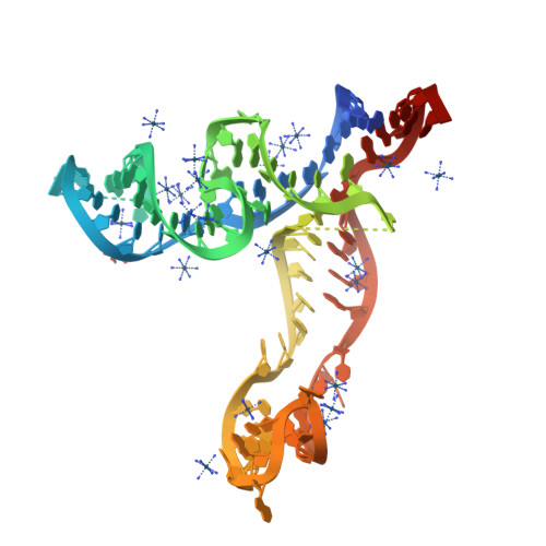
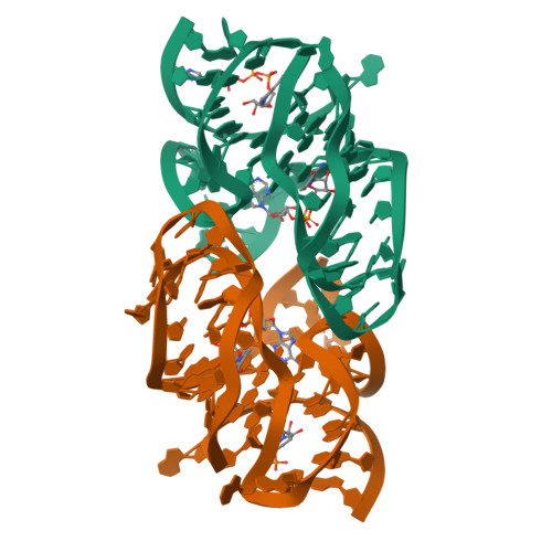
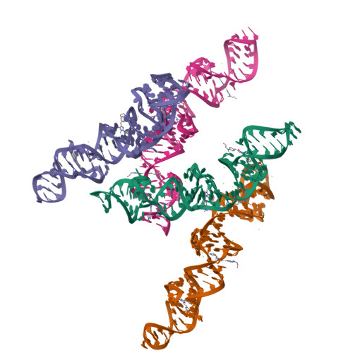
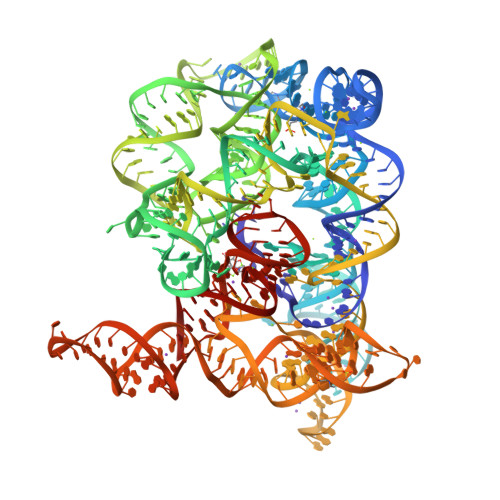
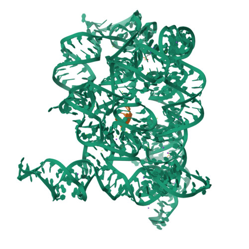
Apo structure of the TPP riboswitch aptamer domain
SM: PG4
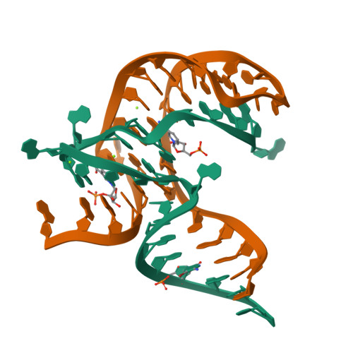
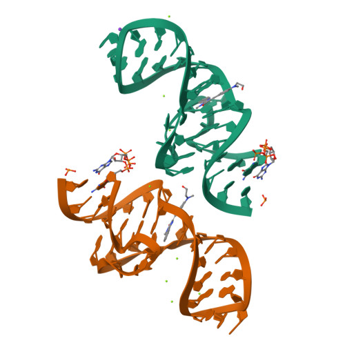
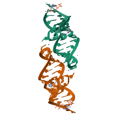
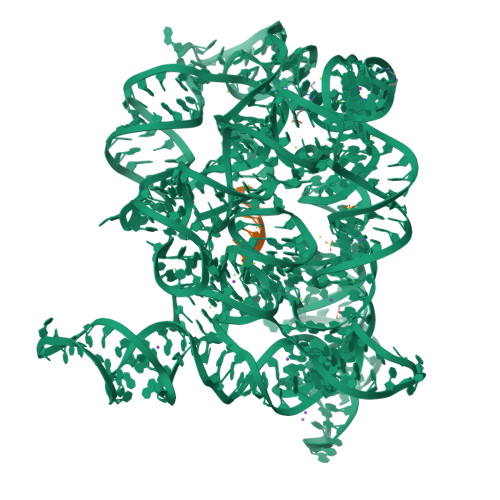
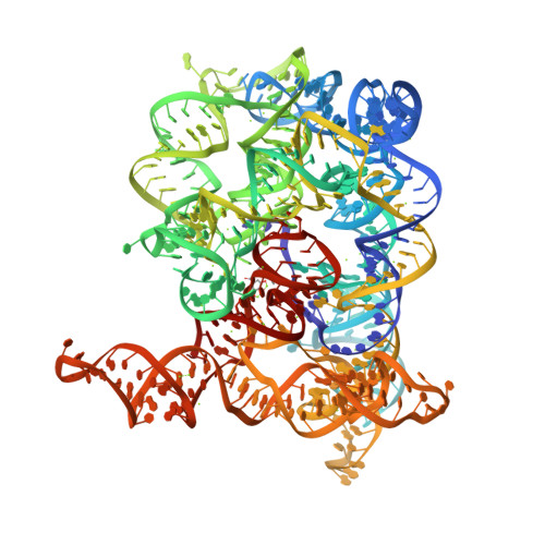
PreQ1-1 (type-1) riboswitch with stacked metabolites and a C10-G34 base pair in the expression platform
SM: PRF PRF PRF PRF PRF PRF
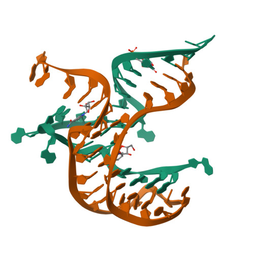
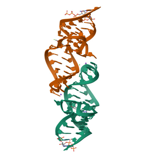
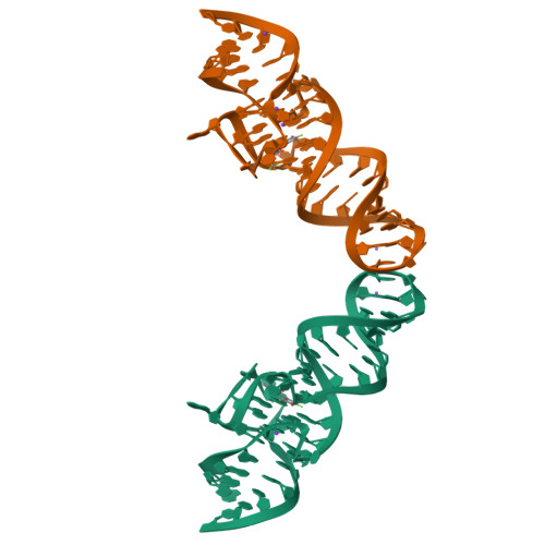
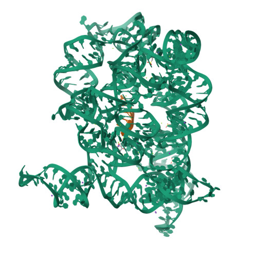
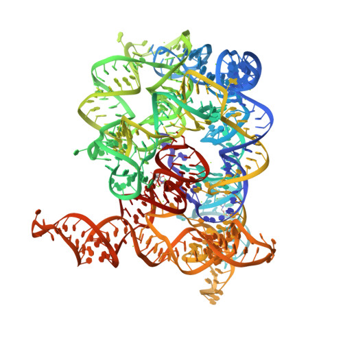
Class I type III preQ1 riboswitch from E. coli
SM: PRF PRF
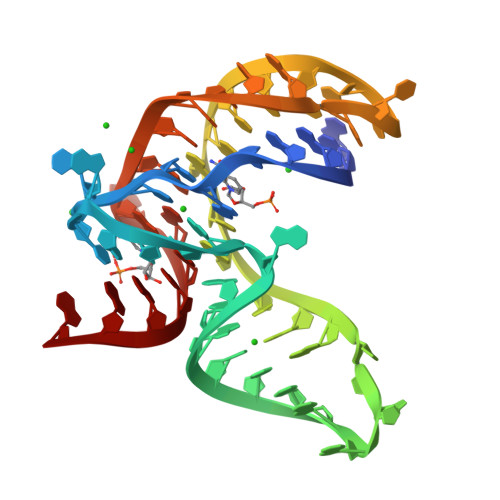
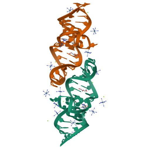
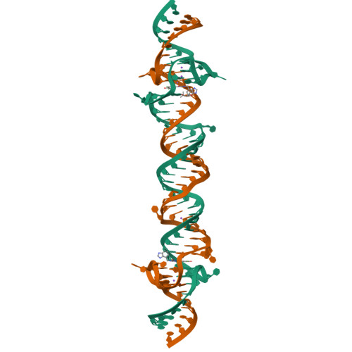
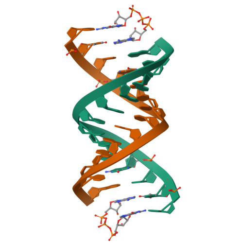
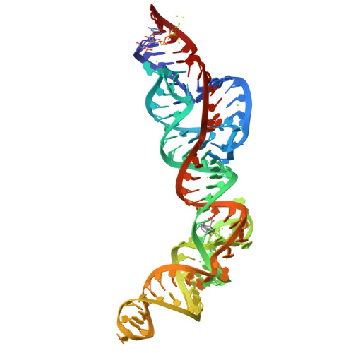
Crystal structure of NAD-II riboswitch (two strands) with NMN
SM: NMN NMN
Crystal structure of NAD-II riboswitch (two strands) with NR
SM: NNR NNR
Crystal structure of NAD-II riboswitch (single strand) with NMN
Crystal structure of NAD-II riboswitch (single strand) with NAD
SM: NAD NAD NAD NMN
A new fluorescent RNA aptamer bound with N618
SM: O2I O2I
A new fluorescent RNA aptamer bound with N
SM: NI4 NI4
A new fluorescent RNA aptamer bound with N565
SM: NJL NJL
A new fluorescent RNA aptamer bound with N571
SM: O00 O00
A new fluorescent RNA aptamer bound with N, iridium hexammine soak
A new fluorescent RNA aptamer_III bound with N
SM: NI4 NI4 NI4 NI4 NI4 NI4
A new fluorescent RNA aptamer bound with N, manganese soak
Crystal structure of NAD-II riboswitch (two strands) with NMN at 1.67 angstrom
Interaction between a fluoroquinolone derivative KG022 and RNAs: effect of base pairs 3' adjacent to the bulge out residues
SM: 53D
The Tet-S1 state of G264A mutated Tetrahymena group I intron with 6nt 3'/5'-exon and 2-aminopurine nucleoside
SM: OJI
Crystal structure of Broccoli aptamer with DFHBI-1T
SM: 2ZY
Crystal structure of Red Broccoli aptamer with OBI
SM: A1EBI A1EBI
Crystal structure of 2'-dG-III riboswitch with 2'-dG
SM: GNG
Crystal structure of 2'-dG-III riboswitch with guanosine
SM: GMP GMP
Structure of Oceanobacillus iheyensis group II intron in the presence of K+, Mg2+ and intronistat B
SM: EPE EPE SPM SPM VTE
Structure of Oceanobacillus iheyensis group II intron in the presence of K+, Mg2+ and ARN25850
SM: EPE EPE SPM VTR
Structure of Oceanobacillus iheyensis group II intron post first step of splicing in the presence of K+, Mg2+ and intronistat B
SM: EPE EPE EPE SPM SPM
Structure of Oceanobacillus iheyensis group II intron in the presence of K+, Mg2+, 5'-exon, and intronistat B after 2h30 soaking
SM: EPE SPM VTE
Structure of Oceanobacillus iheyensis group II intron in the presence of Na+, Mg2+ and intronistat B
SM: GDE
Crystal structure of RNA G2C4 repeats in complex with small synthetic molecule ANP77
SM: W53
RNA duplex bound with GpppA dinucleotide ligand
SM: G3A G3A
GpppA dinucleotide ligand binding to RNA UC template
Solution structure of Risdiplam bound to the RNA duplex formed upon 5'-splice site recognition
SM: Y59
Solution structure of branaplam bound to the RNA duplex formed upon 5'-splice site recognition
SM: Y53
Solution structure of SMN-CX bound to the RNA helix formed upon SMN2 exon7 5'-splice site recognition
SM: YB3
GpppA dinucleotide binding to RNA CU template
RNA duplex bound with GMP and AMP monomers
SM: 5GP 5GP AMP
Structure of Oceanobacillus iheyensis group II intron in the presence of K+, Mg2+ and 5'-exon
SM: EPE
Structure of Oceanobacillus iheyensis group II intron in the presence of K+, Mg2+, 5'-exon, and intronistat B after 1h soaking
SM: VTE
Structure of Oceanobacillus iheyensis group II intron in the presence of K+, Mg2+, 5'-exon, and ARN25850 after 1h soaking
SM: VTR
Structure of Oceanobacillus iheyensis group II intron in the presence of Na+, Mg2+, and ARN25850
Structure of Oceanobacillus iheyensis group II intron in the presence of Li+ and Mg2+
SM: EPE EPE
Structure of Oceanobacillus iheyensis group II intron in the presence of Li+, Mg2+, and intronistat B
Structure of Oceanobacillus iheyensis group II intron in the presence of Li+, Mg2+, and ARN25850
SM: EPE EPE VTR
RNA UU template binding to AMP monomer
SM: AMP AMP AMP
RNA duplex bound with imidazolium bridged GA dinucleotide
SM: WZW WZW WZW WZW
Crystal structure of RNA device 43 truncation mutant 3 (U100C), holo state
SM: TAC TAC TAC TAC
Cryo-EM structure of RNA device 43, holo state
SM: TAC
Class III PreQ1 riboswitch double mutant U8C/A85G
SM: PRF
Structure of Mango III variant aptamer bound to T01-07M-B
SM: W6F
Structure of Mango II aptamer bound to T01-6A
SM: VK0 VK0 VK0
Structure of Mango II aptamer bound to T01-6A-B
SM: VKI
Structure of Mango II variant aptamer bound to T01-6A-B
SM: VLR VLR VLR
Structure of Mango II variant aptamer bound to T01-7M-B
SM: W6F W6F
Structure of Mango II variant2 aptamer bound to T01-6A
Magnesium catalyzed primer extension product with 14mer primer and mixed phosphorothioate activated G monomer
SM: DGP DGP
Cadmium catalyzed primer extension product with 14mer primer and mixed phosphorothioate activated G monomer
Class III PreQ1 riboswitch mutant delta84
Structure of S. odontolytica ZTP riboswitch bound to m-1-pyridinyl-AICA
SM: UG4
SM: A1AD3
Current selection range: to
Filter entries " Experimental resolution " between the two values. Global DB range: 0.6 – 11.2
Filter entries " Molecular weight (compound) " between the two values. Global DB range: 150 – 1000
Filter entries " Deposition date " between the two values. Global DB range: 1986 – 2027
Filter entries " Molecular weight (complex) " between the two values. Global DB range: 0 – 13090
Filter entries " Number of atoms " between the two values. Global DB range: 5 – 70
Filter entries " Number of interacting chains " between the two values. Global DB range: 1 – 4
Filter entries " Pocket size " between the two values. Global DB range: 15.9 – 394.0
Filter entries " SiteScore " between the two values. Global DB range: 0.3 – 1.3
Filter entries " Dscore " between the two values. Global DB range: -0.1 – 1.4
Filter entries " Exposure " between the two values. Global DB range: 0.1 – 1.0
Filter entries " Enclosure " between the two values. Global DB range: 0.3 – 1.0
Filter entries " Hydrophobicity " between the two values. Global DB range: 0.0 – 3.1
Filter entries " Hydrophilicity " between the two values. Global DB range: 0.2 – 2.8
Filter entries " Donor/Acceptor " between the two values. Global DB range: 0.0 – 5.0
Filter entries " Volume " between the two values. Global DB range: 21.2 – 1528.8