Harnessing RIBOnucleic acid - Small molecules Structures
Combine filters to refine your query. Each filter opens a dialog showing the values available in the data.
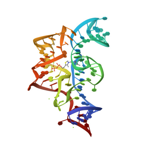
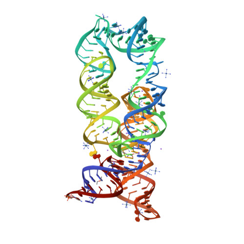
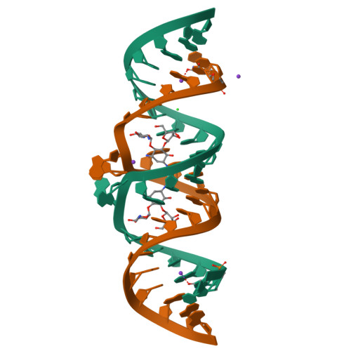
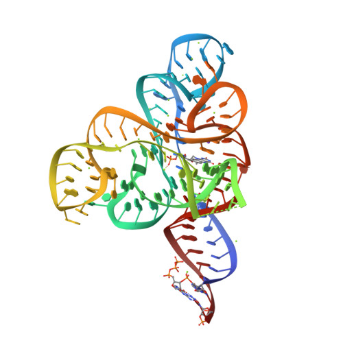
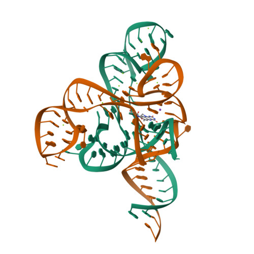
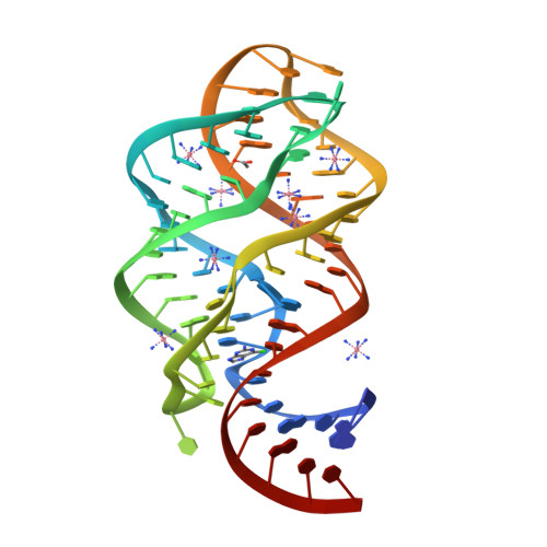
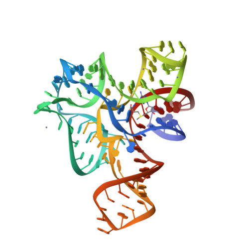
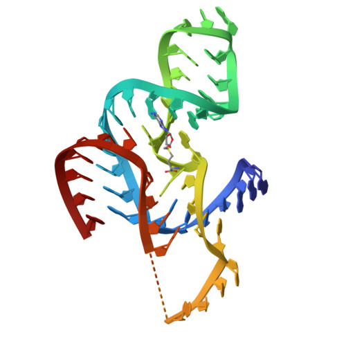
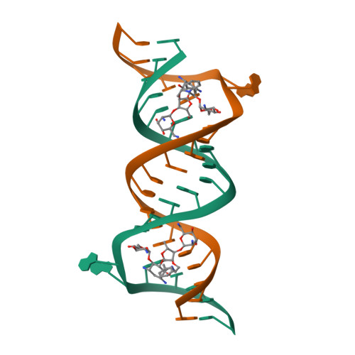
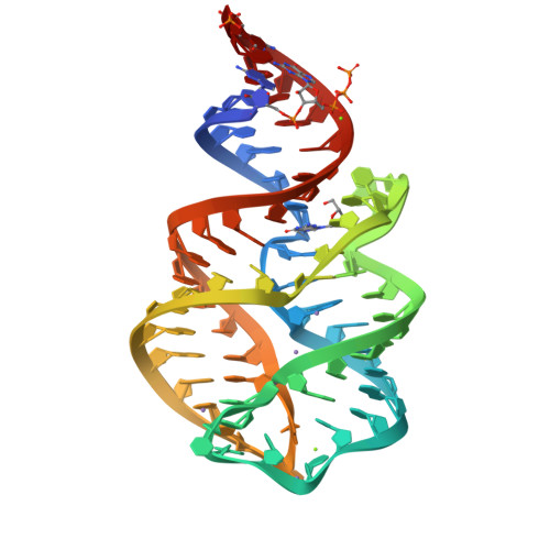
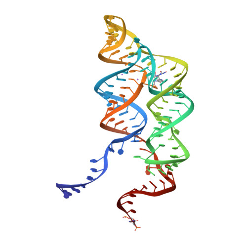
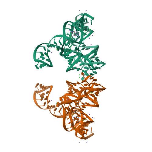
Structure of the thiamine pyrophosphate-specific riboswitch bound to oxythiamine pyrophosphate
SM: D2X D2X
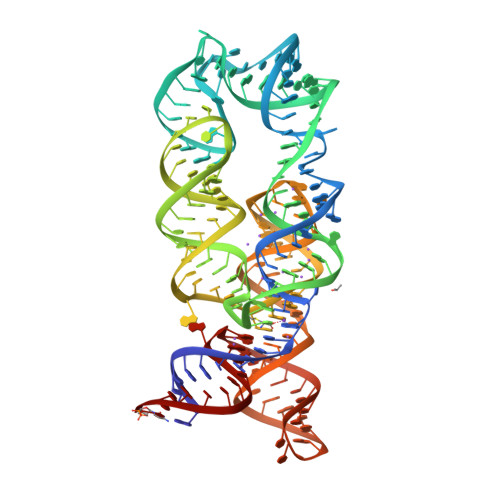
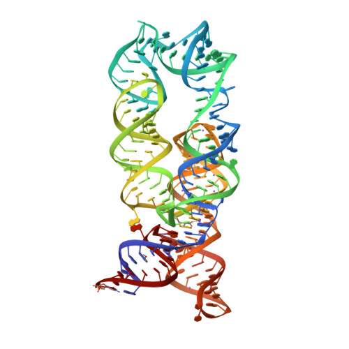
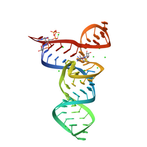
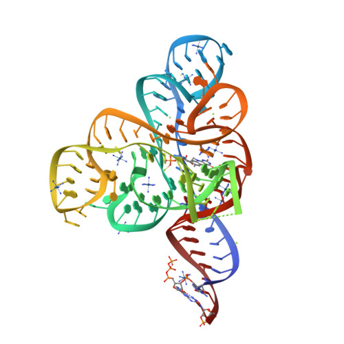
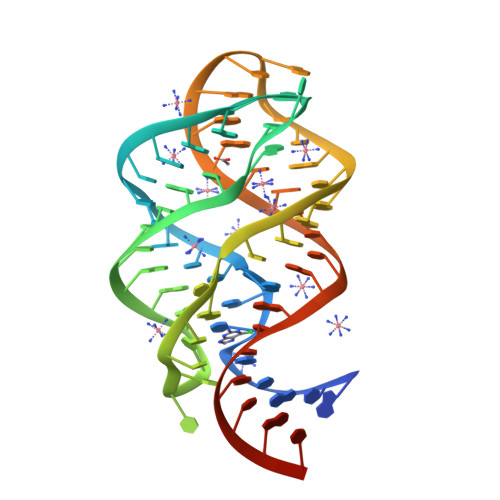
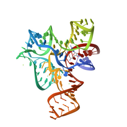
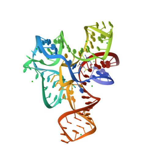
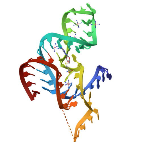
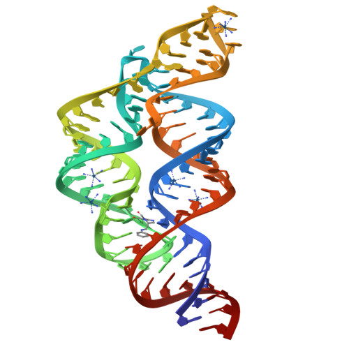
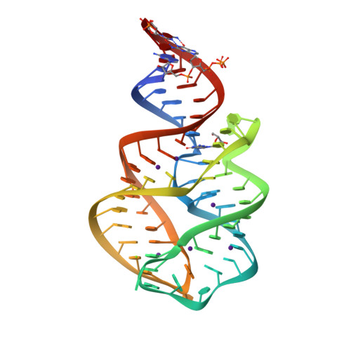
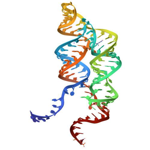
CRYSTAL STRUCTURE OF THE THERMOTOGA MARITIMA LYSINE RIBOSWITCH BOUND TO S-(2-aminoethyl)-L-cysteine
SM: SLZ
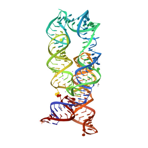
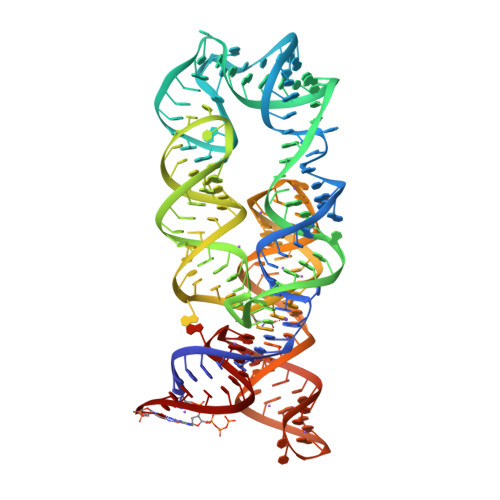
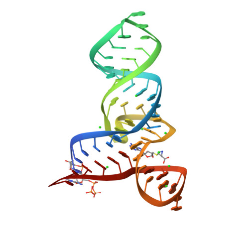
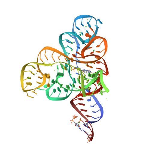
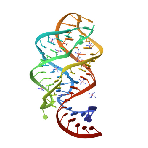
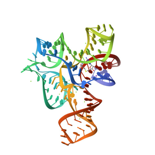
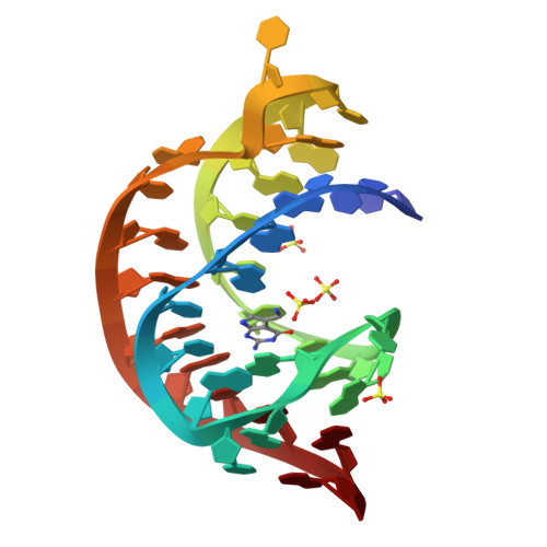
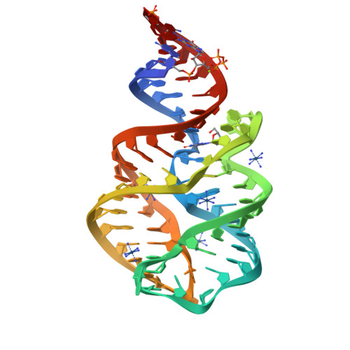
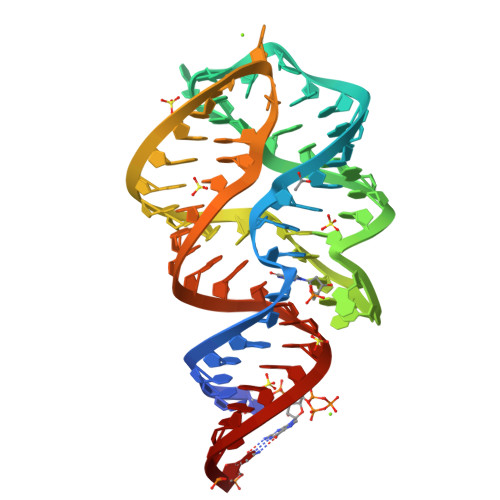
Crystal structure of the Thermotoga maritima lysine riboswitch bound to lysine
SM: 1PE 1PE 1PE
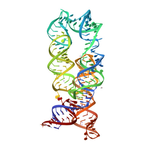
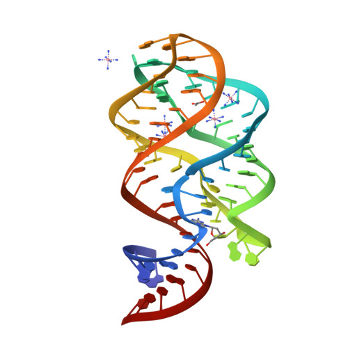
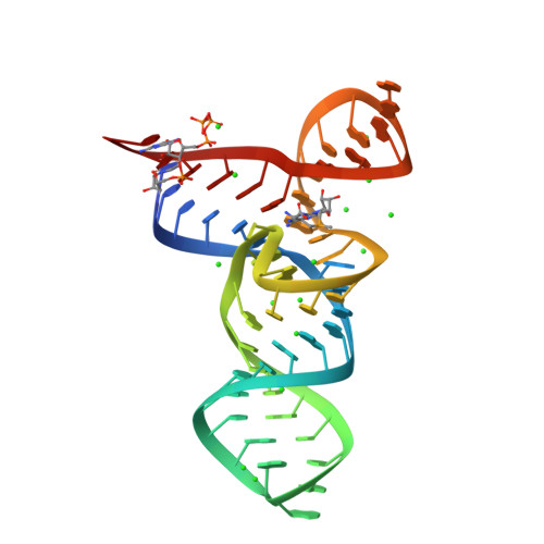
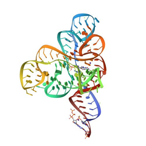
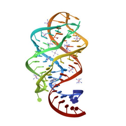
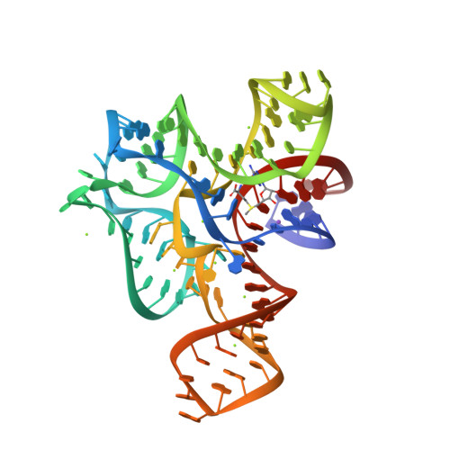
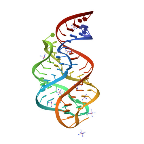
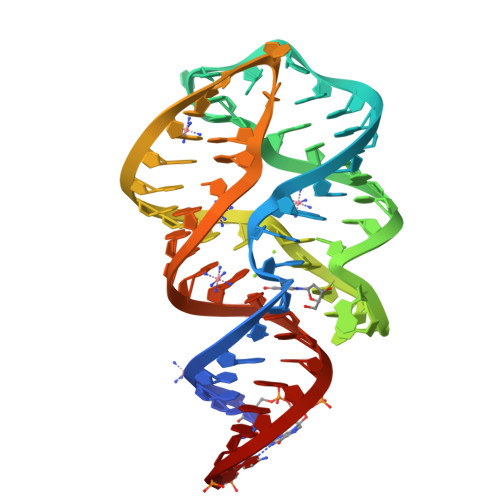
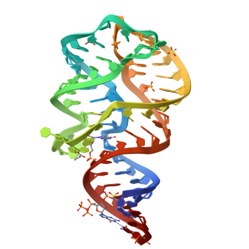
Crystallization of the Thermotoga maritima lysine riboswitch bound to lysine, Cs+ Soak
SM: 1PE
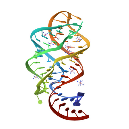
Crystallization of the Thermotoga maritima lysine riboswitch bound to lysine, IRIDIUM HEXAMINE SOAK
Crystallization of the Thermotoga maritima lysine riboswitch bound to homoarginine
SM: 1PE HRG
Crystallization of the Thermotoga maritima lysine riboswitch bound to N6-1-iminoethyl-L-Lysine
SM: IEL
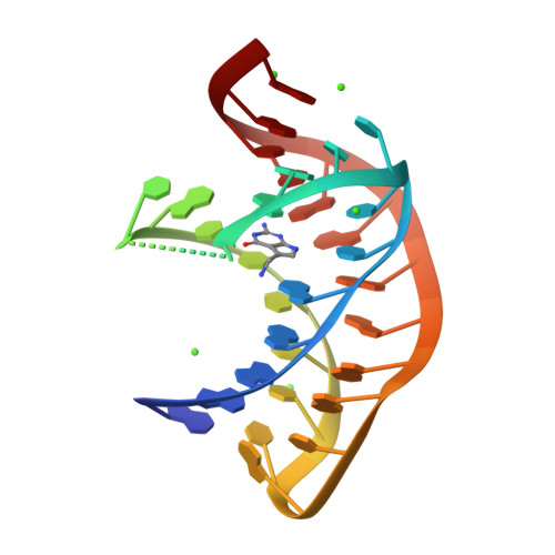
Structure of an RNA-2'-deoxyguanosine complex
SM: GNG GNG
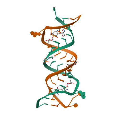
Crystal structure of HIV-1 subtype F DIS extended duplex RNA bound to ribostamycin (U267OMe)
SM: RIO RIO
Crystal Structure of the SMK box (SAM-III) Riboswitch with SAM
SM: SAM
Crystal Structures of the SMK box (SAM-III) Riboswitch with SAH
SM: SAH
Crystal Structures of the SMK box (SAM-III) Riboswitch with Se-SAM
SM: EEM
Crystal structure of the FMN riboswitch bound to FMN
SM: FMN
Crystal structure of the FMN riboswitch bound to FMN, iridium hexamine soak.
Crystal structure of the FMn riboswitch bound to FMN, Ba2+ soak.
Crystal structure of the FMN riboswitch bound to FMN, Cs+ soak.
Crystal structure of the FMN riboswitch bound to FMN, Mn2+ soak.
Crystal structure of the FMN riboswitch bound to FMN, cobalt hexammine soak.
Crystal structure of the FMN riboswitch bound to FMN, split RNA.
Crystal structure of the FMN riboswitch bound to riboflavin.
SM: RBF
Crystal structure of the FMN riboswitch bound to roseoflavin
SM: RS3
Crystal structure of guanine riboswitch C74U mutant bound to 6-chloroguanine
SM: 6GU
Crystal structure of guanine riboswitch bound to 6-O-methylguanine
SM: 6GO
Cocrystal structure of a class-I preQ1 riboswitch
SM: PRF PRF PRF
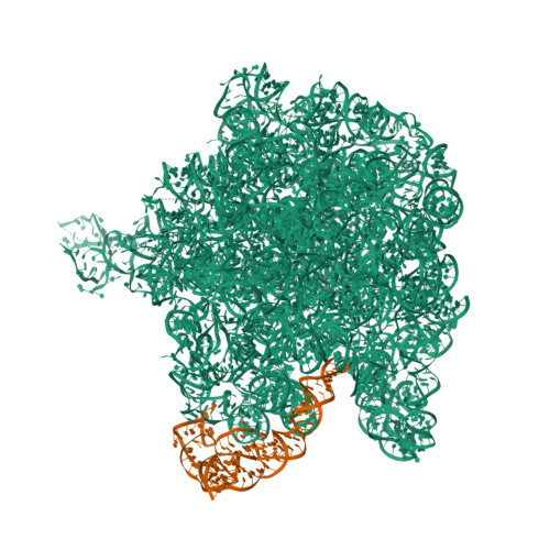
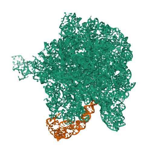
The large ribosomal subunit from Deinococcus radiodurans complexed with Methymycin
SM: MT9
The structural basis for recognition of the preQ0 metabolite by an unusually small riboswitch aptamer domain
SM: PQ0
Guanine riboswitch bound to 6-chloroguanine
Crystal structure of the guanine riboswitch C74U mutant bound to 6-O-methylguanine
Guanine riboswitch A21G,U75C mutant bound to 6-chloroguanine
TteSAM-I riboswitch variant A94GU34C bound to sinefungin
SM: SFG
Crystal structure of the T. tengcongensis SAM-I riboswitch variant U34C/A94G bound with SAH
Crystal structure of T. tencongensis SAM-I riboswitch variant A94G/U34 bound with SAM
Crystal structure of the T. tengcongensis SAM-I riboswitch variant U34C/A94G bound with SAM in manganese chloride
Crystal structure of the T. tengcongensis SAM-I riboswitch variant U34C/A94G mutant A6C/U7G/A87C/U88G bound with SAM
Free-state structural transitions of the SAM-I riboswitch
SAM-I riboswitch from T. tencongensis variant A94G bound with SAM
The structure of the complex of the large ribosomal subunit from D. Radiodurans with the antibiotic lankacidin
SM: LC2
Cocrystal structure of a mutant class-I preQ1 riboswitch
SM: PRF
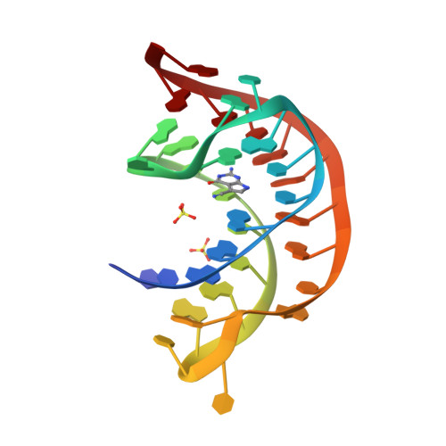
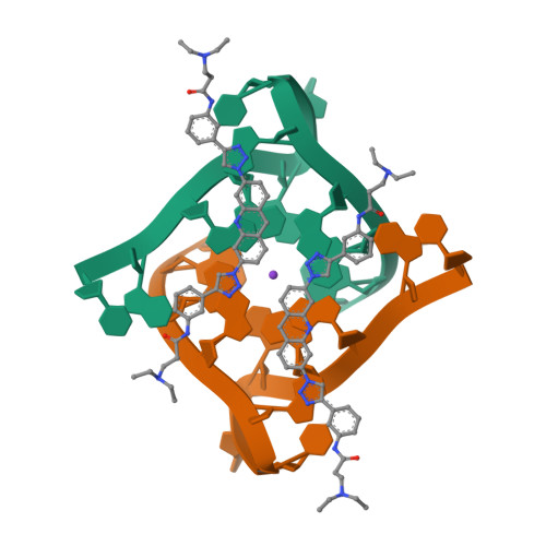
Crystal structure of a telomeric RNA G-quadruplex complexed with an acridine-based ligand.
SM: R14
Structure of the s-adenosylhomocysteine riboswitch at 3.0A
Structure of the S-adenosylhomocysteine riboswitch at 2.18 A
SM: SAH SAH SAH
Structure of a c-di-GMP-II riboswitch from C. acetobutylicum bound to c-di-GMP
SM: C2E C2E C2E C2E
Structural analysis of a class I PreQ1 riboswitch aptamer in the metabolite-bound state
Crystal structure of guanine riboswitch C61U/G37A double mutant bound to thio-guanine
SM: DX4 DX4 DX4 DX4
Crystal structure of the bacterial ribosomal decoding site complexed with an amphiphilic paromomycin O2''-ether analogue
SM: JS6 JS6
The structure of the tetrahydrofolate riboswitch containing a U25C mutation
SM: FFO
Crystal structure of the 2'- Deoxyguanosine riboswitch bound to 2'-deoxyguanosine
Crystal structure of the 2'- deoxyguanosine riboswitch bound to 2'-deoxyguanosine, iridium hexammine soak
Crystal structure of the 2'- Deoxyguanosine riboswitch bound to 2'- Deoxyguanosine, cobalt Hexammine soak
Crystal structure of the 2'- Deoxyguanosine riboswitch bound to 2'- Deoxyguanosine, manganese Soak
Crystal structure of the 2'- Deoxyguanosine riboswitch bound to 2'- Deoxyguanosine, cesium soak
Crystal structure of the 2'- deoxyguanosine riboswitch bound to guanosine
SM: GMP GMP
Crystal structure of the 2'- Deoxyguanosine riboswitch bound to 2'-deoxyguanosine-5'-monophosphate
SM: DGP DGP
Crystal structure of the 2'- Deoxyguanosine riboswitch bound to guanosine-5'-monophosphate
SM: 5GP 5GP
Crystal structure of THF riboswitch, bound with 5-formyl-THF
Crystal structure of THF riboswitch, bound with THF
SM: THF
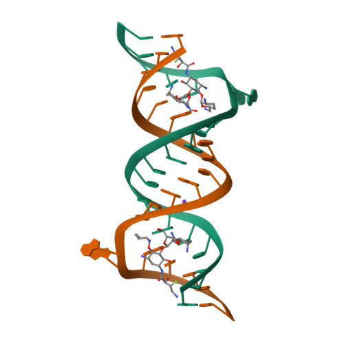
Crystal structure of the bacterial A1408G-mutant and the protozoa cytoplasmic ribosomal decoding site in complex with geneticin
SM: GET GET
Structure of a Riboswitch-like RNA-ligand complex from the Hepatitis C Virus Internal Ribosome Entry Site
SM: SS0
Crystal structure of the bacterial ribosomal decoding site in complex with synthetic aminoglycoside with F-HABA group
SM: SJP SJP
SAM-I riboswitch containing the T. solenopsae Kt-23 in complex with S- adenosyl methionine
Current selection range: to
Filter entries " Experimental resolution " between the two values. Global DB range: 0.6 – 11.2
Filter entries " Molecular weight (compound) " between the two values. Global DB range: 150 – 1000
Filter entries " Deposition date " between the two values. Global DB range: 1986 – 2027
Filter entries " Molecular weight (complex) " between the two values. Global DB range: 0 – 13090
Filter entries " Number of atoms " between the two values. Global DB range: 5 – 70
Filter entries " Number of interacting chains " between the two values. Global DB range: 1 – 4
Filter entries " Pocket size " between the two values. Global DB range: 15.9 – 394.0
Filter entries " SiteScore " between the two values. Global DB range: 0.3 – 1.3
Filter entries " Dscore " between the two values. Global DB range: -0.1 – 1.4
Filter entries " Exposure " between the two values. Global DB range: 0.1 – 1.0
Filter entries " Enclosure " between the two values. Global DB range: 0.3 – 1.0
Filter entries " Hydrophobicity " between the two values. Global DB range: 0.0 – 3.1
Filter entries " Hydrophilicity " between the two values. Global DB range: 0.2 – 2.8
Filter entries " Donor/Acceptor " between the two values. Global DB range: 0.0 – 5.0
Filter entries " Volume " between the two values. Global DB range: 21.2 – 1528.8