HARIBOSS logo

HARIBOSS

Harnessing RIBOnucleic acid - Small molecules Structures

RNA-SM complexes

× Close

Combine filters to refine your query. Each filter opens a dialog showing the values available in the data.


1146 RNA-SM complexes found
Structure of complex 2o3x

2o3x

Crystal Structure of the Prokaryotic Ribosomal Decoding Site Complexed with Paromamine Derivative NB30

SM: N30

Structure of complex 2o3y

2o3y

Crystal Structure of the Homo sapiens Cytoplasmic Ribosomal Decoding Site in Presence of Paromamine Derivative NB30

SM: SPM

Structure of complex 2o43

2o43

Structure of 23S rRNA of the large ribosomal subunit from Deinococcus radiodurans in complex with the macrolide erythromycylamine

SM: ERN

Structure of complex 2o44

2o44

Structure of 23S rRNA of the large ribosomal subunit from Deinococcus radiodurans in complex with the macrolide josamycin

SM: JOS

Structure of complex 2o45

2o45

Structure of the 23S rRNA of the large ribosomal subunit from Deinococcus Radiodurans in complex with the macrolide RU-69874

SM: RU6

Structure of complex 2oe5

2oe5

1.5 A X-ray crystal structure of Apramycin complex with RNA fragment GGCGUCGCUAGUACCG/GGUACUAAAAGUCGCCC containing the human ribosomal decoding A site: RNA construct with 3'-overhang

SM: AM2

Structure of complex 2oe8

2oe8

1.8 A X-ray crystal structure of Apramycin complex with RNA fragment GGGCGUCGCUAGUACC/CGGUACUAAAAGUCGCC containing the human ribosomal decoding A site: RNA construct with 5'-overhang

SM: AM2

Structure of complex 2ogn

2ogn

The crystal structure of the large ribosomal subunit from Deinococcus radiodurans complexed with the pleuromutilin derivative SB-280080

SM: G80

Structure of complex 2ogo

2ogo

The crystal structure of the large ribosomal subunit from Deinococcus radiodurans complexed with the pleuromutilin derivative retapamulin (SB-275833)

SM: G34

Structure of complex 2pwt

2pwt

Crystal structure of the bacterial ribosomal decoding site complexed with aminoglycoside containing the L-HABA group

SM: LHA LHA LHA

Structure of complex 2qwy

2qwy

SAM-II riboswitch bound to S-adenosylmethionine

SM: SAM SAM SAM

Structure of complex 2tra

2tra

RESTRAINED REFINEMENT OF TWO CRYSTALLINE FORMS OF YEAST ASPARTIC ACID AND PHENYLALANINE TRANSFER RNA CRYSTALS

SM: SPM

Structure of complex 2uu9

2uu9

Structure of the Thermus thermophilus 30S ribosomal subunit complexed with a Valine-ASL with cmo5U in position 34 bound to an mRNA with a GUG-codon in the A-site and paromomycin.

SM: PAR

Structure of complex 2uua

2uua

Structure of the Thermus thermophilus 30S ribosomal subunit complexed with a Valine-ASL with cmo5U in position 34 bound to an mRNA with a GUC-codon in the A-site and paromomycin.

SM: PAR

Structure of complex 2uub

2uub

Structure of the Thermus thermophilus 30S ribosomal subunit complexed with a Valine-ASL with cmo5U in position 34 bound to an mRNA with a GUU-codon in the A-site and paromomycin.

SM: PAR

Structure of complex 2uuc

2uuc

Structure of the Thermus thermophilus 30S ribosomal subunit complexed with a Valine-ASL with cmo5U in position 34 bound to an mRNA with a GUA-codon in the A-site and paromomycin.

SM: PAR

Structure of complex 2uxb

2uxb

Crystal structure of an extended tRNA anticodon stem loop in complex with its cognate mRNA GGGU in the context of the Thermus thermophilus 30S subunit.

SM: PAR

Structure of complex 2uxc

2uxc

Crystal structure of an extended tRNA anticodon stem loop in complex with its cognate mRNA UCGU in the context of the Thermus thermophilus 30S subunit.

SM: PAR

Structure of complex 2uxd

2uxd

Crystal structure of an extended tRNA anticodon stem loop in complex with its cognate mRNA CGGG in the context of the Thermus thermophilus 30S subunit.

SM: PAR

Structure of complex 2vqe

2vqe

Modified uridines with C5-methylene substituents at the first position of the tRNA anticodon stabilize U-G wobble pairing during decoding

SM: PAR

Structure of complex 2vqf

2vqf

Modified uridines with C5-methylene substituents at the first position of the tRNA anticodon stabilize U-G wobble pairing during decoding

SM: PAR

Structure of complex 2ydh

2ydh

Crystal structure of the SAM-I riboswitch A94G U34 G18U G19U variant in complex with SAM

SM: SAM

Structure of complex 2ygh

2ygh

SAM-I riboswitch with a G2nA mutation in the Kink turn in complex with S-adenosylmethionine

SM: SAM

Structure of complex 2yie

2yie

Crystal structure of a F. nucleatum FMN riboswitch bound to FMN

SM: FMN

Structure of complex 38jc

38jc

RNA polymerase ribozyme 85h34 replication complex, consensus structure.

SM: APC

Structure of complex 38jd

38jd

RNA polymerase ribozyme 85h34 replication complex, subclass 2 structure

SM: APC

Structure of complex 38je

38je

RNA polymerase ribozyme 85h34 replication complex, subclass 7 structure

SM: APC

Structure of complex 3b4a

3b4a

T. tengcongensis glmS ribozyme with G40A mutation, bound to glucosamine-6-phosphate

SM: GLP

Structure of complex 3b4b

3b4b

T. tengcongensis glmS ribozyme with G40A mutation, bound to glucosamine-6-phosphate and a substrate RNA with a 2'5'-phosphodiester linkage

SM: GLP

Structure of complex 3b4c

3b4c

T. tengcongensis glmS ribozyme bound to glucosamine-6-phosphate and a substrate RNA with a 2'5'-phosphodiester linkage

SM: GLP

Structure of complex 3bnq

3bnq

Crystal Structure of the Homo sapiens Mitochondrial Ribosomal Decoding Site in the Presence of SrCl2 (A1555G mutant, Br-derivative)

SM: PAR

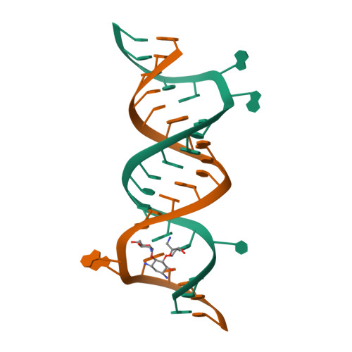
Structure of complex 3bnr

3bnr

Crystal Structure of the Homo sapiens Mitochondrial Ribosomal Decoding Site in the presence of nonspecifically bound paromomycin (A1555G mutant, Br-derivative)

SM: PAR

Structure of complex 3cc4

3cc4

Co-crystal Structure of Anisomycin Bound to the 50S Ribosomal Subunit

SM: ANM

Structure of complex 3cxc

3cxc

The structure of an enhanced oxazolidinone inhibitor bound to the 50S ribosomal subunit of H. marismortui

SM: SLD

Structure of complex 3d2g

3d2g

Structural basis of thiamine pyrophosphate analogues binding to the eukaryotic riboswitch

SM: TPP TPP

Structure of complex 3d2v

3d2v

Structure of the eukaryotic TPP-specific riboswitch bound to the antibacterial compound pyrithiamine pyrophosphate

SM: PYI PYI

Structure of complex 3d2x

3d2x

Structure of the thiamine pyrophosphate-specific riboswitch bound to oxythiamine pyrophosphate

SM: D2X D2X

Structure of complex 3dig

3dig

CRYSTAL STRUCTURE OF THE THERMOTOGA MARITIMA LYSINE RIBOSWITCH BOUND TO S-(2-aminoethyl)-L-cysteine

SM: SLZ

Structure of complex 3dil

3dil

Crystal structure of the Thermotoga maritima lysine riboswitch bound to lysine

SM: 1PE 1PE 1PE

Structure of complex 3dim

3dim

Crystallization of the Thermotoga maritima lysine riboswitch bound to lysine, Cs+ Soak

SM: 1PE

Structure of complex 3dio

3dio

Crystallization of the Thermotoga maritima lysine riboswitch bound to lysine, IRIDIUM HEXAMINE SOAK

SM: 1PE

Structure of complex 3diq

3diq

Crystallization of the Thermotoga maritima lysine riboswitch bound to homoarginine

SM: 1PE HRG

Structure of complex 3dir

3dir

Crystallization of the Thermotoga maritima lysine riboswitch bound to N6-1-iminoethyl-L-Lysine

SM: IEL

Structure of complex 3dll

3dll

The oxazolidinone antibiotics perturb the ribosomal peptidyl-transferase center and effect tRNA positioning

SM: ZLD

Structure of complex 3ds7

3ds7

Structure of an RNA-2'-deoxyguanosine complex

SM: GNG GNG

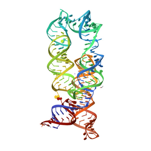
Structure of complex 3e5c

3e5c

Crystal Structure of the SMK box (SAM-III) Riboswitch with SAM
SM: SAM

Structure of complex 3e5e

3e5e

Crystal Structures of the SMK box (SAM-III) Riboswitch with SAH

SM: SAH

Structure of complex 3e5f

3e5f

Crystal Structures of the SMK box (SAM-III) Riboswitch with Se-SAM

SM: EEM

Structure of complex 3egz

3egz

Crystal structure of an in vitro evolved tetracycline aptamer and artificial riboswitch

SM: CTC

Structure of complex 3f2q

3f2q

Crystal structure of the FMN riboswitch bound to FMN

SM: FMN

Structure of complex 3f2t

3f2t

Crystal structure of the FMN riboswitch bound to FMN, iridium hexamine soak.

SM: FMN

Structure of complex 3f2w

3f2w

Crystal structure of the FMn riboswitch bound to FMN, Ba2+ soak.

SM: FMN

Structure of complex 3f2x

3f2x

Crystal structure of the FMN riboswitch bound to FMN, Cs+ soak.

SM: FMN

Structure of complex 3f2y

3f2y

Crystal structure of the FMN riboswitch bound to FMN, Mn2+ soak.

SM: FMN

Structure of complex 3f30

3f30

Crystal structure of the FMN riboswitch bound to FMN, cobalt hexammine soak.

SM: FMN

Structure of complex 3f4e

3f4e

Crystal structure of the FMN riboswitch bound to FMN, split RNA.

SM: FMN

Structure of complex 3f4g

3f4g

Crystal structure of the FMN riboswitch bound to riboflavin.

SM: RBF

Structure of complex 3f4h

3f4h

Crystal structure of the FMN riboswitch bound to roseoflavin

SM: RS3

Structure of complex 3fo4

3fo4

Crystal structure of guanine riboswitch C74U mutant bound to 6-chloroguanine

SM: 6GU

Structure of complex 3fo6

3fo6

Crystal structure of guanine riboswitch bound to 6-O-methylguanine

SM: 6GO