Harnessing RIBOnucleic acid - Small molecules Structures
Combine filters to refine your query. Each filter opens a dialog showing the values available in the data.
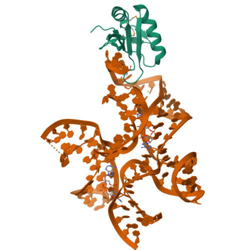
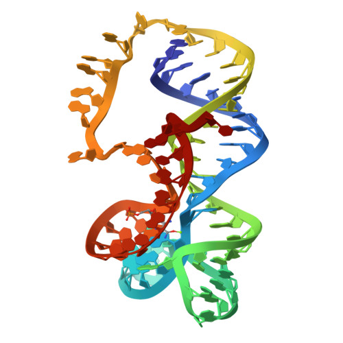
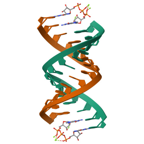
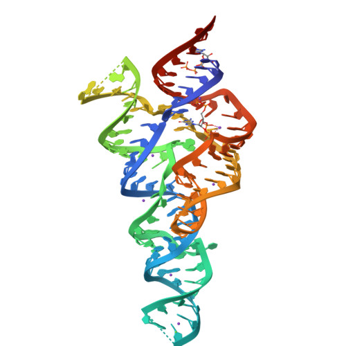
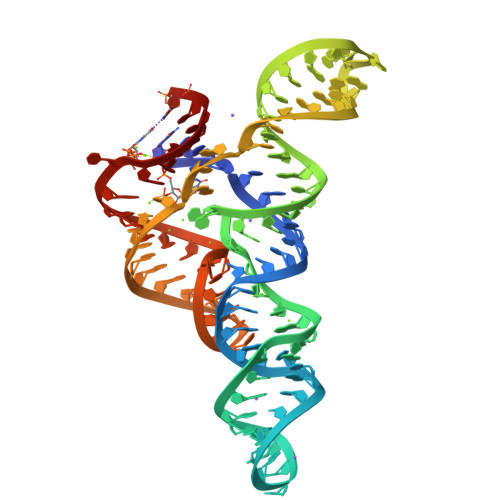
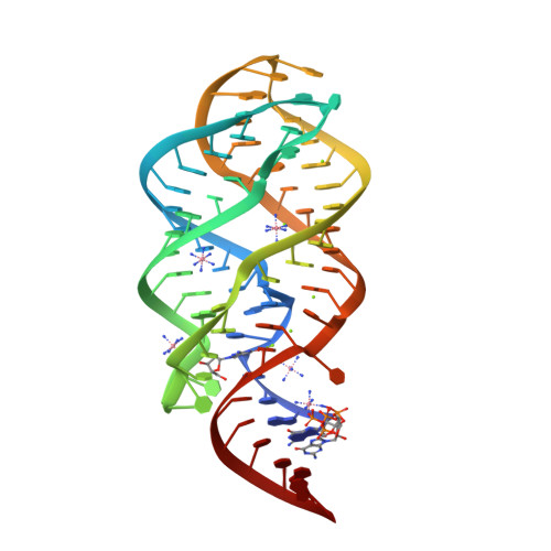
Crystal structure of Bacillus subtilis cyclic-di-AMP riboswitch ydaO
SM: 2BA 2BA
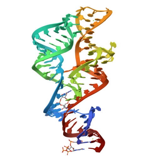
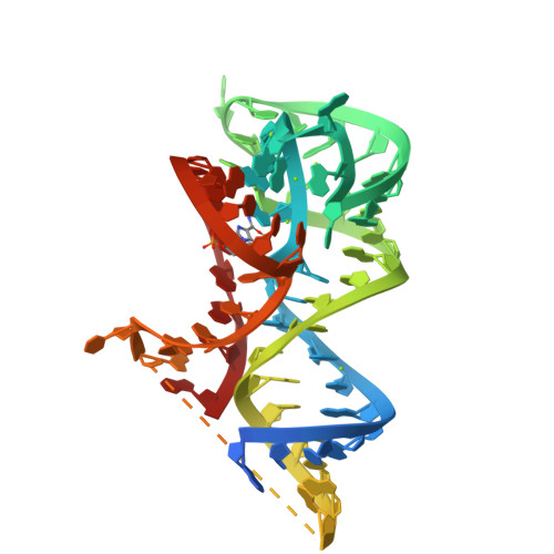
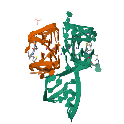
3',3'-cGAMP riboswitch bound with 3',3'-cGAMP
SM: 4BW 4BW
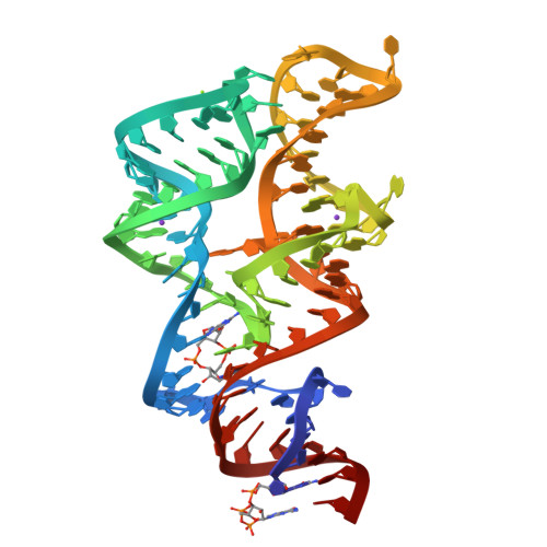
3',3'-cGAMP riboswitch bound with c-di-GMP
SM: C2E C2E
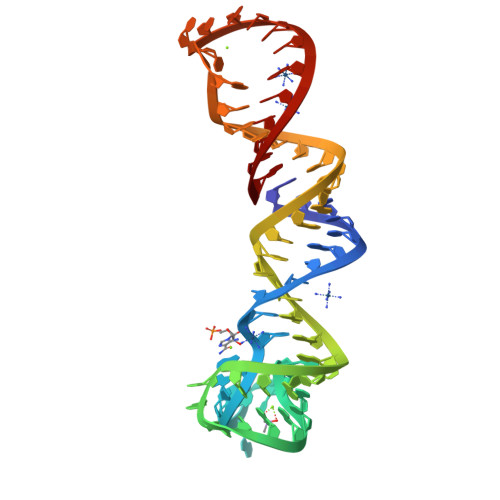
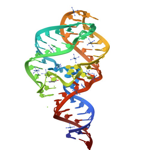
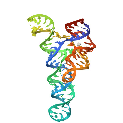
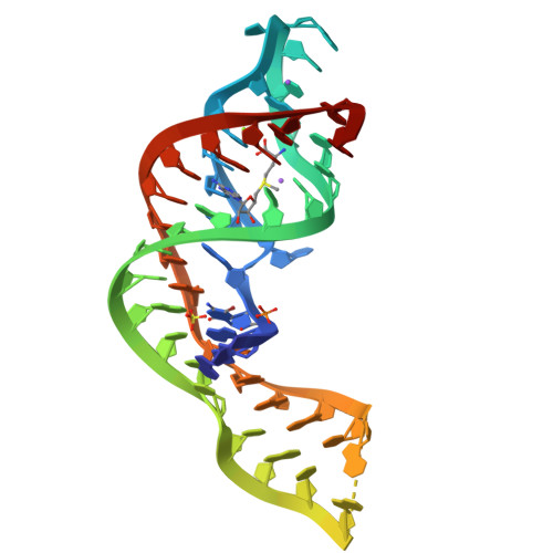
Crystal structure of the ZMP riboswitch at 1.80 angstrom
SM: AMZ
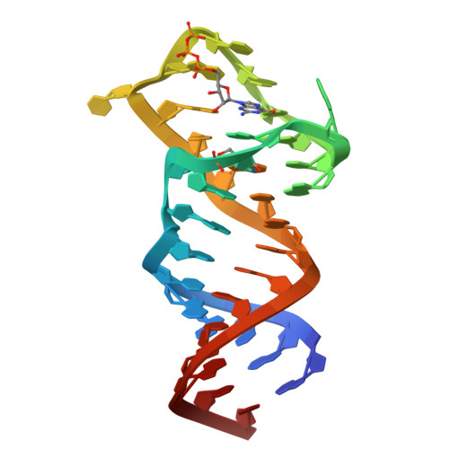
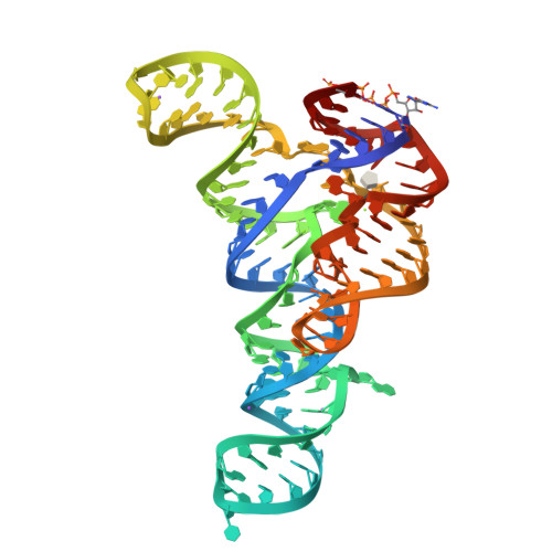
The structure of A pfI Riboswitch Bound to ZMP
SM: AMZ AMZ
Fusobacterium ulcerans ZTP riboswitch bound to ZMP
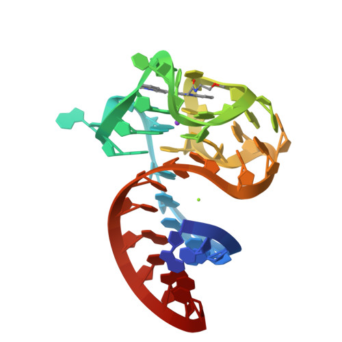
Structure of a 5-hydroxytryptophan aptamer
SM: 4PQ
Solution NMR structure of the GTP binding Class II RNA aptamer-ligand-complex containing a protonated adenine nucleotide with a highly shifted pKa.
SM: GTP
RNA primer-template complex with guanosine dinucleotide ligand G(5')ppp(5')G
SM: GP3 GP3
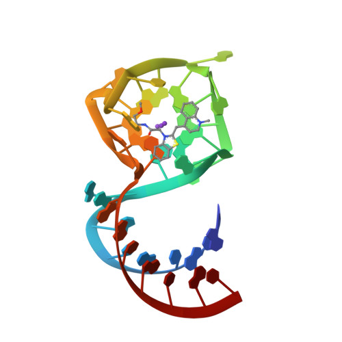
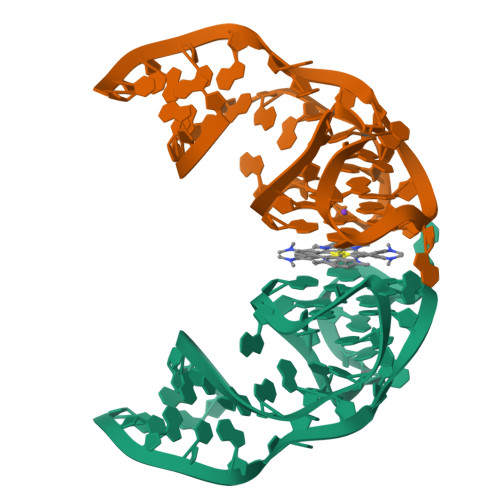
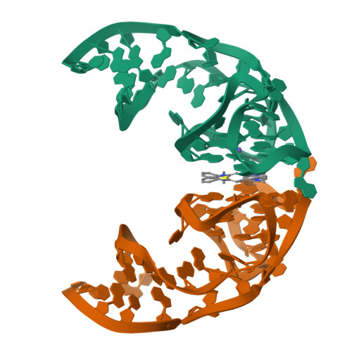
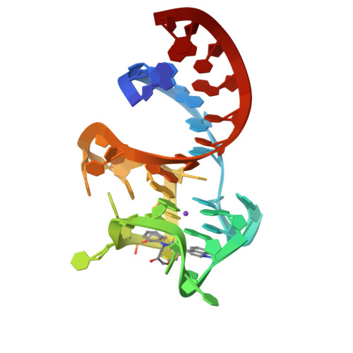
Co-crystal structure of the fluorogenic RNA Mango
SM: 74G 74G
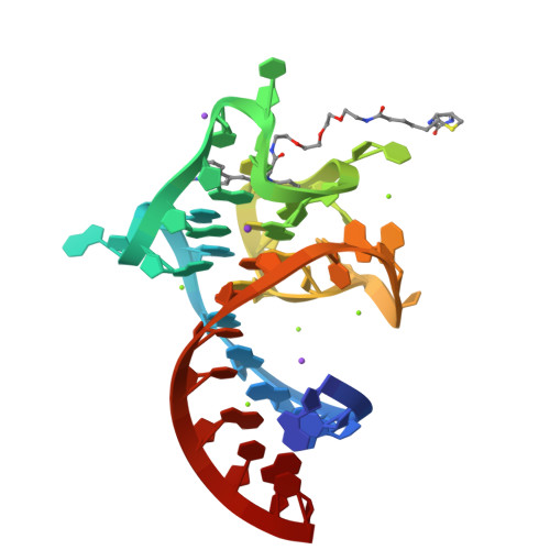
Crystal Structure of the Mango-II Fluorescent Aptamer Bound to TO1-Biotin
SM: EKJ EKJ EKJ
Crystal Structure of the Mango-II Fluorescent Aptamer Bound to TO3-Biotin
SM: EKM EKM
Crystal Structure of the Mango-II-A22U Fluorescent Aptamer Bound to TO1-Biotin
G96A mutant of the PRPP riboswitch from T. mathranii bound to ppGpp
SM: G4P G4P G4P G4P GTP
PRPP riboswitch from T. mathranii bound to PRPP
SM: PRP PRP
PRPP Riboswitch bound to PRPP, native structure
SM: PRP
ppGpp Riboswitch bound to ppGpp, native structure
SM: G4P G4P
Crystal structure of the Corn aptamer in complex with ThT
SM: TFX
Crystal structure of the Corn aptamer in complex with TO
SM: J0D
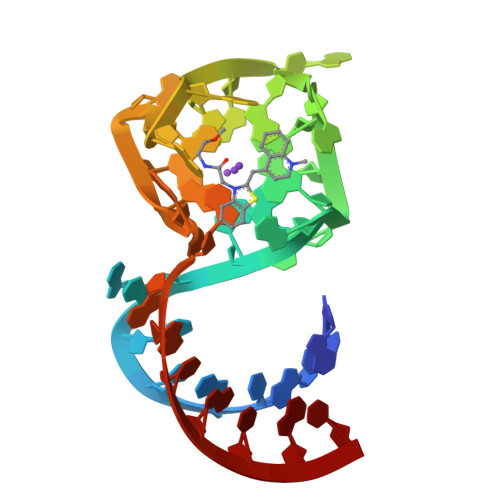
Structure of the iMango-III aptamer bound to TO1-Biotin
SM: EKJ EKJ SPM
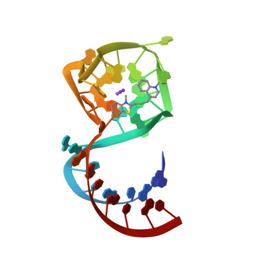
Structure of the Mango-III (A10U) aptamer bound to TO1-Biotin
SM: HZG HZG HZG HZG
SM: HZD
Crystal structure of the metY SAM V riboswitch
SM: SAM
The structure of the SAM/SAH-binding riboswitch.
SM: SAH
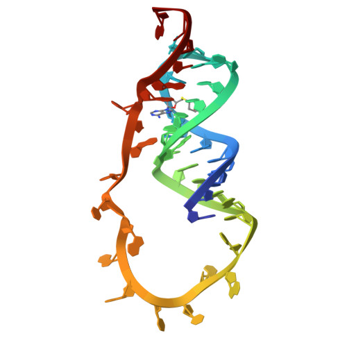
The structure of the G. violaceus guanidine II riboswitch P2 stem-loop with arcaine
SM: FXQ
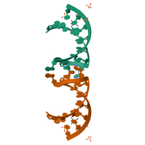
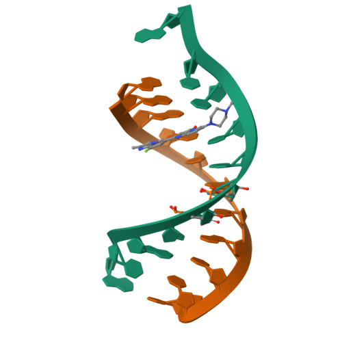
Solution structure of the RNA duplex formed by the 5'-end of U1snRNA and the 5'-splice site of SMN2 exon7 in complex with the SMN-C5 splicing modifier
SM: GDZ
Structural basis for 2'-deoxyguanosine recognition by the 2'-dG-II class of riboswitches
SM: GNG
Current selection range: to
Filter entries " Experimental resolution " between the two values. Global DB range: 0.6 – 11.2
Filter entries " Molecular weight (compound) " between the two values. Global DB range: 150 – 1000
Filter entries " Deposition date " between the two values. Global DB range: 1986 – 2027
Filter entries " Molecular weight (complex) " between the two values. Global DB range: 0 – 13090
Filter entries " Number of atoms " between the two values. Global DB range: 5 – 70
Filter entries " Number of interacting chains " between the two values. Global DB range: 1 – 4
Filter entries " Pocket size " between the two values. Global DB range: 15.9 – 394.0
Filter entries " SiteScore " between the two values. Global DB range: 0.3 – 1.3
Filter entries " Dscore " between the two values. Global DB range: -0.1 – 1.4
Filter entries " Exposure " between the two values. Global DB range: 0.1 – 1.0
Filter entries " Enclosure " between the two values. Global DB range: 0.3 – 1.0
Filter entries " Hydrophobicity " between the two values. Global DB range: 0.0 – 3.1
Filter entries " Hydrophilicity " between the two values. Global DB range: 0.2 – 2.8
Filter entries " Donor/Acceptor " between the two values. Global DB range: 0.0 – 5.0
Filter entries " Volume " between the two values. Global DB range: 21.2 – 1528.8